SUPERFICIAL STRUCTURES
Skin
Skin of upper limb on anterior aspect is more thick, with less hairs and more sensitive. Skin of posterior aspect shows more hairs, thick and less sensitive. Tension lines are present on skin transversely placed on flexor surface and verticle on dorsal surface. Palmar skin is thick and firm for gripping.
Superficial fascia
It forms subcutaneous layer of fibrofatty tissue.
Deep fascia
It divide forearm into anterior and posterior compartments. It forms bicipital aponeurosis, flexor and extensor retinaculum, palmar aponeurosis.
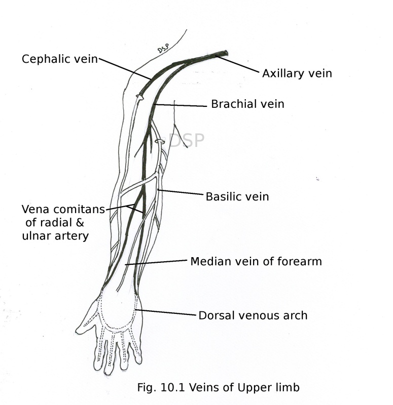
Veins
Veins of upper limb are superficial and deep.
Superficial veins runs in superficial fascia.
Superficial veins are cephalic, basilic, median cubital, median vein.
Cephalic vein
It begins on dorsal aspect of hand on radial side from lateral aspect of dorsal venous arch. It goes upwards on lateral aspect of forearm near elbow joined by median cubital vein. Goes upwards on lateral aspect of arm. Then it pierces deep fascia and drains into axillary vein.
Basilic vein
It begins on dorsal aspect of hand on ulnar side from medial aspect of dorsal venous arch. It goes upwards on medial aspect of forearm near elbow at medial epicondyle of humerus connected to cephalic vein by median cubital vein. Goes upwards on medial aspect of arm. Then it pierces deep fascia and join venae comitantes of brachial artery to form axillary vein. It is used for cardiac catheterisation.
Median cubital vein
It is present on anterior aspect of elbow joint. It joins cephalic and basilic vein at elbow joint. It goes upward and medially. It also gives branch to join with venae comitantes of brachial artery. It is easy accessible vein for withdrawal of venous blood and other procedures like intravenous injection.
Deep veins of upper limb runs with arteries as venae comitantes.
Skin
Skin of upper limb on anterior aspect is more thick, with less hairs and more sensitive. Skin of posterior aspect shows more hairs, thick and less sensitive. Tension lines are present on skin transversely placed on flexor surface and verticle on dorsal surface. Palmar skin is thick and firm for gripping.
Superficial fascia
It forms subcutaneous layer of fibrofatty tissue.
Deep fascia
It divide forearm into anterior and posterior compartments. It forms bicipital aponeurosis, flexor and extensor retinaculum, palmar aponeurosis.
Veins
Veins of upper limb are superficial and deep.
Superficial veins runs in superficial fascia.
Superficial veins are cephalic, basilic, median cubital, median vein.
Cephalic vein
It begins on dorsal aspect of hand on radial side from lateral aspect of dorsal venous arch. It goes upwards on lateral aspect of forearm near elbow joined by median cubital vein. Goes upwards on lateral aspect of arm. Then it pierces deep fascia and drains into axillary vein.
Basilic vein
It begins on dorsal aspect of hand on ulnar side from medial aspect of dorsal venous arch. It goes upwards on medial aspect of forearm near elbow at medial epicondyle of humerus connected to cephalic vein by median cubital vein. Goes upwards on medial aspect of arm. Then it pierces deep fascia and join venae comitantes of brachial artery to form axillary vein. It is used for cardiac catheterisation.
Median cubital vein
It is present on anterior aspect of elbow joint. It joins cephalic and basilic vein at elbow joint. It goes upward and medially. It also gives branch to join with venae comitantes of brachial artery. It is easy accessible vein for withdrawal of venous blood and other procedures like intravenous injection.
Deep veins of upper limb runs with arteries as venae comitantes.
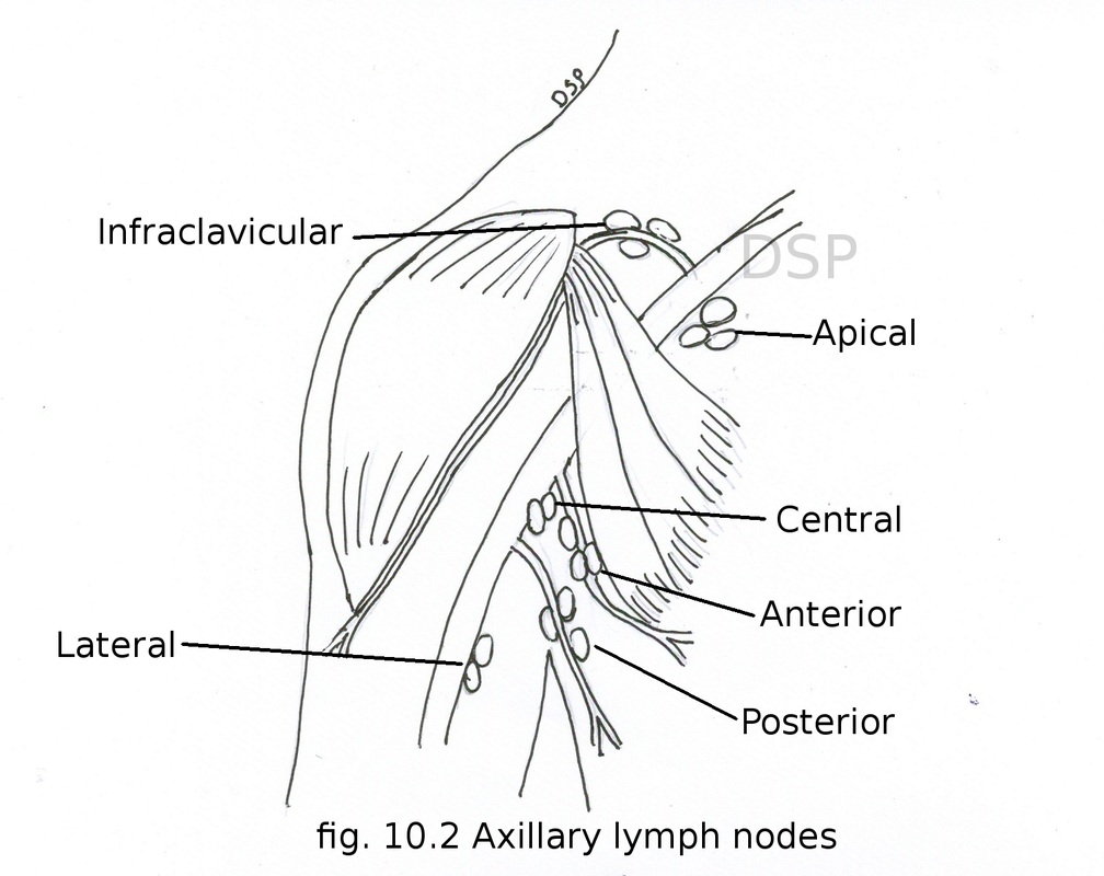
Lymphatic drainage
There are two sets of lymph vessels superficial and deep. These runs alongwith nerves and vessels.
Superficial lymphatics runs alongwith cephalic and basilic veins. Supratrochlear lymph nodes present in relation to basilic vein which drains into lateral group axillary nodes. Infraclavicular nodes in relation to cephalic vein which drains into apical group axillary nodes. Deep lymphatics runs alongwith radial, ulnar and brachial artery. Finally drains into lateral group of axillary nodes.
Axillary lymph nodes
There are 20-30 axillary lymph nodes in axilla. These are divided into lateral, anterior, posterior, central and apical. Axillary nodes drains breast, upper limb and trunk above umbilicus. Lateral group lie posteromedial to axillary vein. It drains complete upper limb except vessels alongwith cephalic vein. Anterior group lie in relation to lateral thoracic vein at inferior border of pectoralis minor. It drains area anterolateral body wall above umbilicus and breast. Posterior group lie along subscapular vessels on posterior wall of axilla. It drains dorsal aspect of trunk upto iliac crest.
Central group lie in axillary fat. It receives lymph from all other groups. It drains in to apical group. Apical group lie near apex of axilla medial to axillary vein. It receives lymph from lymphatics with cephalic vein (upper limb) and upper margin of breast and other axillary lymph nodes. It drains after forming subclavian trunk in to junction of subclavian vein and internal jugular vein. On left side it drains into thoracic duct.
Applied anatomy: Axillary lymph nodes may enlarge in infection, carcinoma (mammary gland) of area of drainage by them.
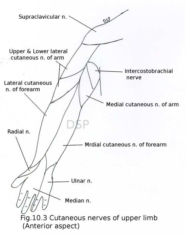
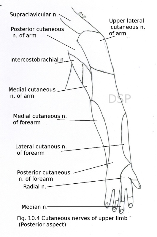
Cutaneous innervation of upper limb
Pectoral region upper part and shoulder region shows nerve supply from supraclavicular nerves. Pectoral region remaining part shows innervation from anterior cutaneous branches of upper six intercostal nerves, lateral cutaneous branches of 3rd to 6th intercostal nerves.
Arm anterior part shows innervation from intercosto-brachial nerve, medial cutaneous nerve of arm, upper and lower lateral cutaneous nerve of arm. Posterior part of arm shows innervation from posterior cutaneous nerve of arm, posterior cutaneous nerve of forearm.
Forearm shows nerve supply from medial and lateral cutaneous nerve of forearm, posterior cutaneous nerve of forearm.
Dorsal surface of hand medial part and medial one and half digits shows nerve supply from dorsal branch of ulnar nerve. Lateral part of dorsum and lateral three and half digits from superficial branch of radial nerve.
Palmar surface of palm shows nerve supply from palmar branch of ulnar nerve and median nerve. Medial one and half digits shows nerve supply from branch of ulnar nerve. Lateral side of palm with lateral three and half digits shows nerve supply from median nerve.