Front of leg and Dorsum of foot
Deep fascia of leg
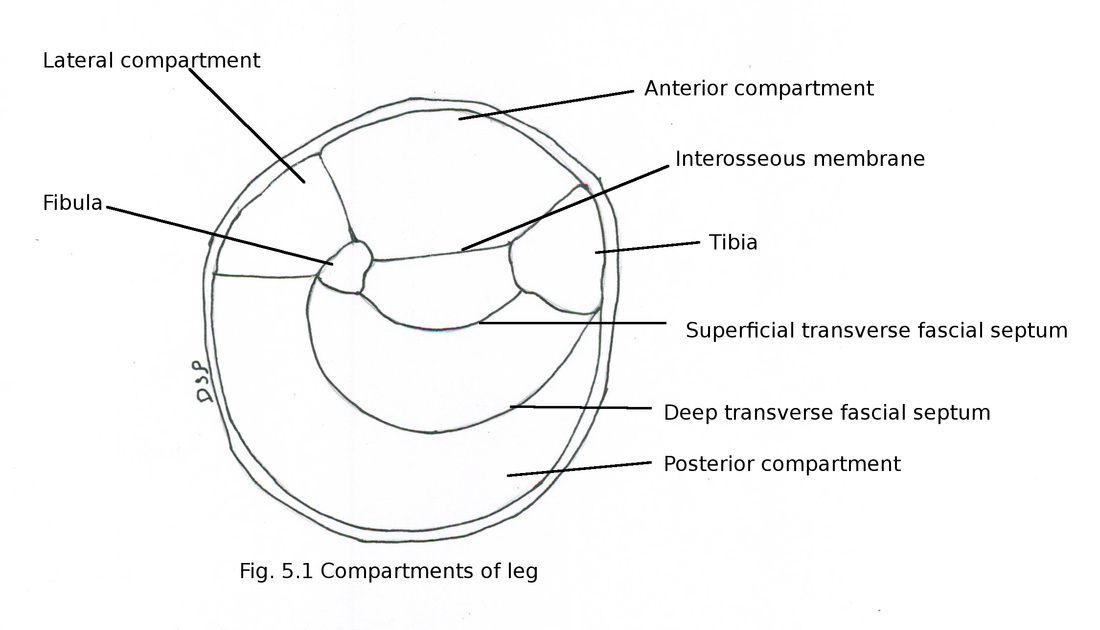
Deep fascia of leg is thick and forms a covering around leg. It is continuous with fascia latae and show attachment on margin of patella, ligamentum patellae, medial and lateral condyles of tibia, tibial tuberosity and head of fibula. Posteriorly over the surface of popliteal fossa it is pierced by small saphenous vein and sural nerve. It forms anterior and posterior intermuscular septum which shows attachment on anterior and posterior margins of fibula. Interosseous membrane shows attachment on interosseous border of tibia and fibula. Upper margin of interosseous membrane shows of gap through which anterior tibial artery passes to anterior compartment. Through a gap present near lower part passes perforating branch of peroneal artery. Two inter muscular septum and interosseous membrane divide leg into anterior, posterior and lateral compartments. Posterior compartment of leg again divided into superficial and deep parts by superficial and deep transverse fascial septum. Deep transverse fascial septum shows attachment on medial margin of tibia and posterior margin of fibula.
Deep fascia of leg
Deep fascia of leg is thick and forms a covering around leg. It is continuous with fascia latae and show attachment on margin of patella, ligamentum patellae, medial and lateral condyles of tibia, tibial tuberosity and head of fibula. Posteriorly over the surface of popliteal fossa it is pierced by small saphenous vein and sural nerve. It forms anterior and posterior intermuscular septum which shows attachment on anterior and posterior margins of fibula. Interosseous membrane shows attachment on interosseous border of tibia and fibula. Upper margin of interosseous membrane shows of gap through which anterior tibial artery passes to anterior compartment. Through a gap present near lower part passes perforating branch of peroneal artery. Two inter muscular septum and interosseous membrane divide leg into anterior, posterior and lateral compartments. Posterior compartment of leg again divided into superficial and deep parts by superficial and deep transverse fascial septum. Deep transverse fascial septum shows attachment on medial margin of tibia and posterior margin of fibula.
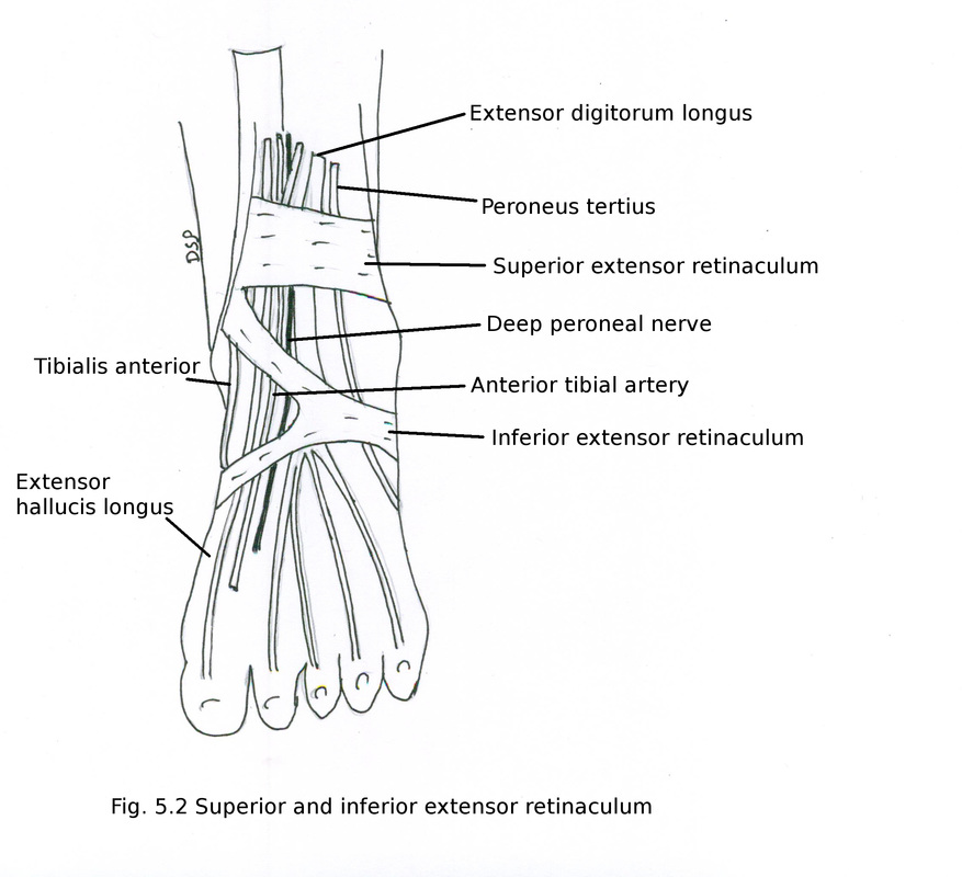
Near lower part of leg on anterior aspect and on dorsum of foot deep fascia get modified into superior and inferior extensor retinaculum.
Superior extensor retinaculum
It is a band of deep fascia present on anterior aspect of lower part of leg just above ankle joint.
Attachments : Medially it shows attachment on anterior border of tibia in its lower part and laterally it shows attachment on anterior border of fibula in its lower part. It enclose tendon of tibialis anterior muscle and structures passing deep to it are extensor hallucis longus, deep peroneal nerve, anterior tibial artery, extensor digitorum longus and peroneus tertius.
Inferior extensor retinaculum
It is a Y shaped band of deep fascia present in front of ankle joint and proximal part of dorsum of foot. It shows a stem which lies laterally and two parts upper and lower band which lie medially.
Attachments : Laterally stem of inferior extensor retinaculum shows attachment on upper surface of calcaneum on its anterior part in front of sulcus calcanei. Upper band goes medially shows attachment on anterior border of medial malleolus. Interior band goes medially over extensor tendons present on dorsum of foot and fuse with plantar aponeurosis.
Structures in relation with inferior extensor retinaculum
Fibres from stem forms a loop around tendons of peroneus tertius and extensor digitorum longus. Upper band enclose tendons of tibialis anterior, extensor hallucis longus. while anterior tibial artery with deep peroneal nerve lies deep to it. Lower band shows tibialis anterior, extensor hallucis longus tendon, dorsalis pedis artery and deep peroneal nerve deep to it.
Functions : It keeps the structures in position present on anterior aspect of leg and dorsum of foot.
Front of leg or Extensor compartment or Anterior compartment of leg
Anterior compartment of leg is bounded by
Anteriorly : Deep fascia of leg
Posteriorly : Interosseous membrane
Medially : Lateral surface of shaft of tibia
Laterally : Anterior intermuscular septum and extensor surface of fibula
Contents
Muscles : Tibialis anterior, Extensor hallucis longus, Extensor digitorum longus, Peroneus tertius
Vessels : Anterior tibial artery
Nerve : Deep peroneal nerve
Muscles :
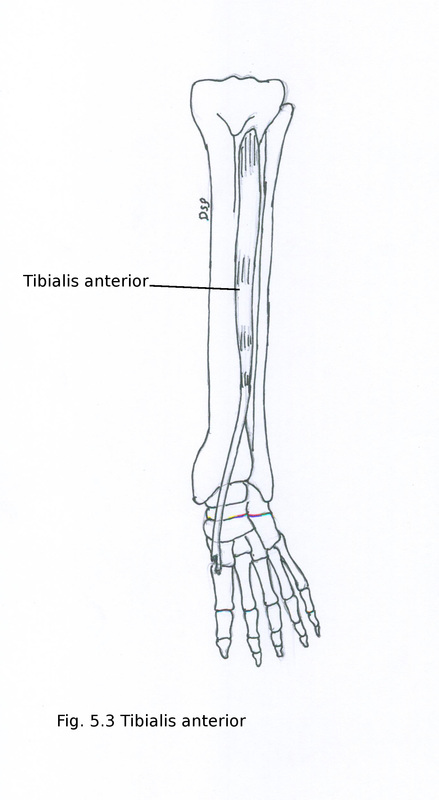
Tibialis anterior
It is a multipennate muscle and spindle shaped.
Origin : It shows origin from upper two part part of lateral surface of tibia, part of lateral condyle tibia, adjacent part of interosseous membrane, deep surface of deep fascia, anterior intermuscular septum.
Insertion : Below in lower one third part of leg this muscle forms a tendon. Tendon passes through superior and inferior extensor retinaculum. It shows insertion on medial and inferior surface of medial cuneiform bone, adjacent surface of first metatarsal bone.
Nerve supply : It receives nerve supply from deep peroneal nerve L4, L5.
Action : Dorsiflexion of foot at ankle joint. Inversion of foot at midtarsal and subtalar jonts . Support medial longitudinal arch from above.
Anterior compartment of leg is bounded by
Anteriorly : Deep fascia of leg
Posteriorly : Interosseous membrane
Medially : Lateral surface of shaft of tibia
Laterally : Anterior intermuscular septum and extensor surface of fibula
Contents
Muscles : Tibialis anterior, Extensor hallucis longus, Extensor digitorum longus, Peroneus tertius
Vessels : Anterior tibial artery
Nerve : Deep peroneal nerve
Muscles :
Tibialis anterior
It is a multipennate muscle and spindle shaped.
Origin : It shows origin from upper two part part of lateral surface of tibia, part of lateral condyle tibia, adjacent part of interosseous membrane, deep surface of deep fascia, anterior intermuscular septum.
Insertion : Below in lower one third part of leg this muscle forms a tendon. Tendon passes through superior and inferior extensor retinaculum. It shows insertion on medial and inferior surface of medial cuneiform bone, adjacent surface of first metatarsal bone.
Nerve supply : It receives nerve supply from deep peroneal nerve L4, L5.
Action : Dorsiflexion of foot at ankle joint. Inversion of foot at midtarsal and subtalar jonts . Support medial longitudinal arch from above.
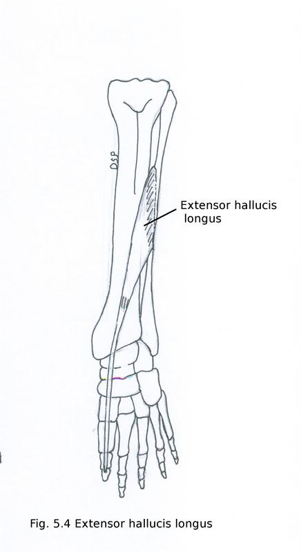
Extensor hallucis longus
It is a unipennate muscle.
Origin : It shows origin from middle two fourth part of medial surface of fibula medial to origin of extensor digitorum longus, adjacent part of interosseous membrane.
Insertion : Below in lower part of leg this muscle forms a tendon. Tendon passes deep to superior and inferior extensor retinaculum. After crossing anterior tibial vessels from lateral to medial side. It shows insertion on dorsal aspect of base of distal phalanx of great toe.
Nerve supply : It receives nerve supply from deep peroneal nerve L5.
Action : Dorsi flexion of great toe and extension of phalanges of great toe.
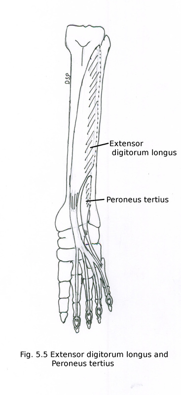
Extensor digitorum longus
Origin : It shows origin from upper three fourth part of medial surface of fibula, from a small strip on lateral condyle of tibia, adjacent part of interosseous membrane, deep surface of deep fascia and anterior intermuscular septum.
Insertion : Below in lower one third part of leg this muscle forms a tendon. Tendon passes deep to superior extensor retinaculum and within loop of inferior extensor retinaculum. Splits into four slips After crossing anterior tibial vessels from lateral to medial side. It splits into for slips shows insertion on lateral four toes. Tendons of second, third, fourth joined laterally at metatarsophalangeal joint by extensor digitorum brevis forming dorsal digital expansion. The dorsal digital expansion shows attachment of lumbricals and interosseous muscles. Each dorsal digital expansion three parts a central and two lateral slips. Central slip shows attachment on base of middle phalanx, two lateral slips shows attachment on base of terminal phalanx on dorsal aspect.
Nerve supply : It receives nerve supply from deep peroneal nerve L5, S1.
Action : Dorsi flexion of ankle alongwith other extensor muscles. Extension of lateral four toes. Help in tightening of plantar aponeurosis.
Peroneus tertius
Origin : It shows origin from lower one fourth part of medial surface of fibula in line with origin of extensor digitorum longus, adjacent interosseous membrane and anterior intermuscular septum.
Insertion : Below tendon passes deep to superior extensor retinaculum and stem of inferior extensor retinaculum. Then shows insertion on base of fifth metatarsal near its base.
Nerve supply : It receives nerve supply from deep peroneal nerve L5, S1.
Action : Dorsi flexion of ankle and eversion of foot.
Vessels
Anterior tibial artery
It is a branch of popliteal artery arises near lower border of popliteus muscle in back of leg. Artery goes to anterior compartment by passing between two heads of tibialis posterior above upper margin of interosseous membrane. It goes downward over anterior surface of interosseous membrane alongwith two venae comitantes. In lower part of leg lies midway between medial and lateral malleolus. On dorsum of foot continue as dorsalis pedis artery. In upper one third part of leg it lies between tibialis anterior and extensor digitorum longus, in middle one third lies between tibialis anterior and extensor hallucis longus, in lower one third lies between extensor digitorum longus and extensor hallucis longus. It is crosssed by tendon of extensor hallucis longus from lateral to medial side.
Branches :
1) Anterior tibial recurrent artery : It ascends upwards and anastomose with circumflex fibular artery and genicular branches of popliteal artery. It takes part in anastomosis around knee joint.
2) Posterior tibial recurrent artery : It arises from anterior tibial artery before its entry in anterior compartment. Anastomoses with inferior genicular branches of popliteal artery.
3) Muscular arteries : It supply adjacent muscles.
4) Lateral malleolar artery : It goes laterally towards lateral malleolus to take part in anastomosis around ankle joint. It anastomoses with perforating branches of posterior tibial and ascending branches of lateral tarsal artery.
5) Medial malleolar artery : It goes medially towards medial malleolus to take part in anastomosis around ankle joint. It anastomoses with branches of posterior tibial and medial plantar artery.
Nerve :
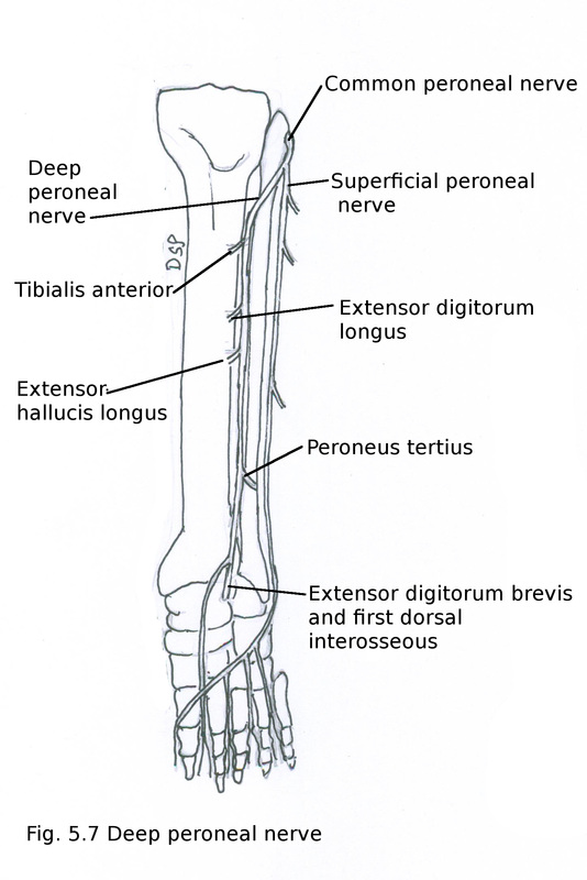
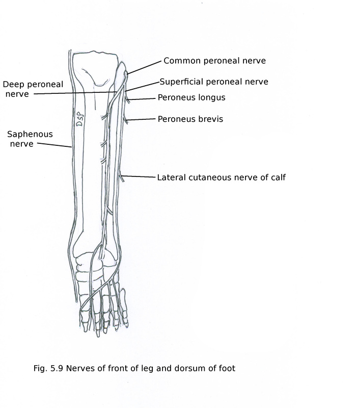
Deep peroneal nerve
It is a branch of common peroneal nerve. It arise near neck of fibula. It goes to anterior compartment by piercing anterior intermuscular septum. It runs in anterior compartment alongwith anterior tibial vessels. It goes down deep to superior and inferior extensor retinaculum. Goes to dorsum of foot and gives out medial and lateral terminal branches. It lies lateral to anterior tibial vessels. Lateral terminal branch forms a pseudo-ganglion deep to extensor digitorum brevis. Branches from ganglion supply extensor digitorum brevis, tarsal and metatarsal joints. Medial terminal branch gives a cutaneous branch to supply skin of first inter-digital cleft and first dorsal interosseous muscle.
Branches :
1) Muscular branches : It supply muscles of anterior compartment of leg tibialis anterior, extensor hallucis longus, extensor digitorum longus, peroneus tertius. Extensor digitorum brevis on dorsum of foot and first dorsal interosseous muscle.
2) Cutaneous branches : It supply skin of first inter-digital cleft.
3) Articular branches : It supply ankle joint.
Applied anatomy: compartment syndrome occurs when there is excessive pressure in anterior compartment due to accumulation of fluid or swelling. It occurs after exercise or injury. Arterial pulsation disappears in dorsalis pedis artery, loss of sensation over first inter-digital cleft. Dorsiflexion of ankle and extension of toes get affected.
Dorsum of foot
Skin and superficial fascia
Subcutaneous layer of skin shows superficial veins. Superficial fascia shows cutaneous nerves and lymphatics.
Superficial veins
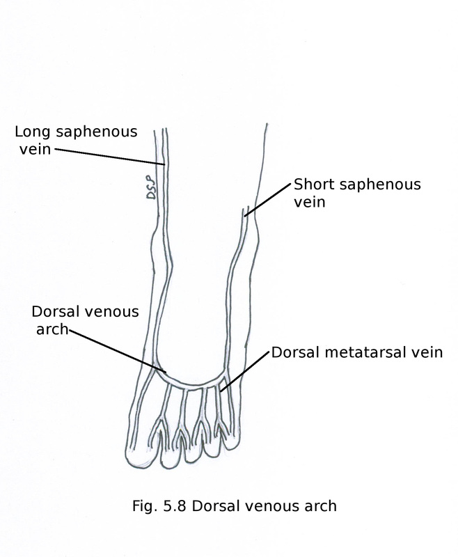
Superficial veins of foot are
1) Dorsal venous arch : It is present on dorsal aspect of foot convex distally in relation with proximal aspect of metatarsal bones. It receives blood from for dorsal metatarsal vein. Its tributaries proximal and distal perforating veins drains blood from plantar aspect of foot.
2) Great saphenous vein : It is continuation of dorsal venous arch medially. Medial marginal vein joins with it on medial side.
3) Small saphenous vein : It is continuation of dorsal venous arch laterally. Lateral marginal vein drains in on lateral side.
4) Dorsal digital veins : Two dorsal digital veins present in relation with each toe. These two join and form dorsal metatarsal vein. Dorsal metatarsal vein drains into dorsal venous arch.
Skin and superficial fascia
Subcutaneous layer of skin shows superficial veins. Superficial fascia shows cutaneous nerves and lymphatics.
Superficial veins
Superficial veins of foot are
1) Dorsal venous arch : It is present on dorsal aspect of foot convex distally in relation with proximal aspect of metatarsal bones. It receives blood from for dorsal metatarsal vein. Its tributaries proximal and distal perforating veins drains blood from plantar aspect of foot.
2) Great saphenous vein : It is continuation of dorsal venous arch medially. Medial marginal vein joins with it on medial side.
3) Small saphenous vein : It is continuation of dorsal venous arch laterally. Lateral marginal vein drains in on lateral side.
4) Dorsal digital veins : Two dorsal digital veins present in relation with each toe. These two join and form dorsal metatarsal vein. Dorsal metatarsal vein drains into dorsal venous arch.
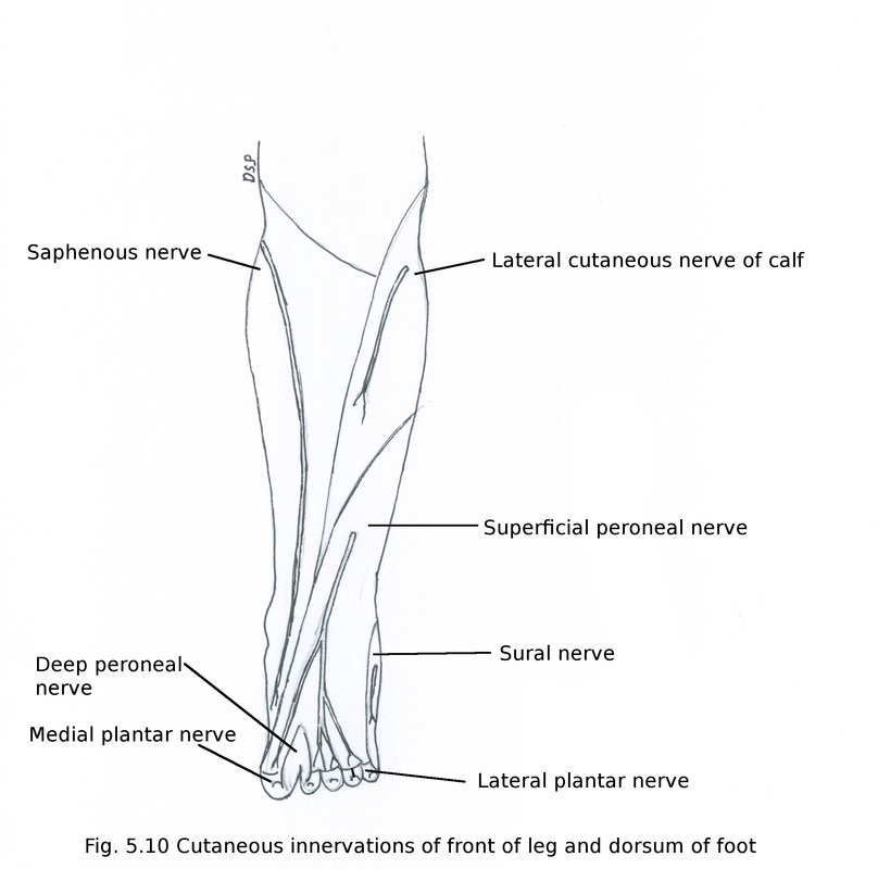
Cutaneous nerves
1) Superficial peroneal nerve (L4, L5, S1) : It is a branch of common peroneal nerve. It arises near neck of fibula. It goes downward and pierce deep fascia of leg near lower one third part. Here it supply lower one third part of leg. It divides into medial and lateral branches. Medial branch supply medial side of great toe and adjacent side of second and third toe. Lateral branch divides into two and supply skin of cleft between third and fourth toes, fourth and fifth toes.
2) Deep peroneal nerve : It supply skin of cleft between first and second toe.
3) Sural nerve : It supply skin of lateral margin of foot and fifth toe.
4) Saphenous nerve : It supply skin of medial margin of foot on dorsum upto head of first metatarsal bone.
Lymphatics
Superficial lymphatics of dorsum drains into vertical groups of superficial inguinal lymph nodes via lymph vessels which runs along with great saphenous vein medially and into popliteal lymph nodes via lymph vessels alongwith short saphenous vein laterally. Deep lymphatic vessels runs along with anterior and posterior tibial vessels, dorsalis pedis artery to drain into popliteal lymph nodes.
Structures present on dorsum of foot
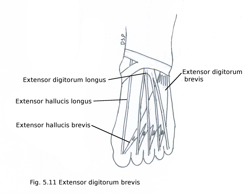
Extrinsic tendons from anterior compartment of leg are tibialis anterior, extensor hallucis longus, extensor digitorum longus, peroneus tertius. Intrinsic muscles present on dorsum are extensor digitorum brevis muscle.
Extensor digitorum brevis
Origin : It shows origin from superior surface of calcaneum from its anterior part and from stem of inferior extensor retinaculum.
Insertion : Muscle divides into four tendons. Medial tendon shows insertion on base of proximal phalanx of great toe also known as extensor hallucis brevis. Remaining three tendons joins with tendons of extensor digitorum longus on lateral side forming dorsal digital expansion of second, third and fourth toes respectively. It shows insertion on base of middle and terminal phalanx of respective toe.
Nerve supply : It receives nerve supply from deep peroneal nerve from its lateral terminal branch.
Action : It helps in dorsiflexion of medial four toes.
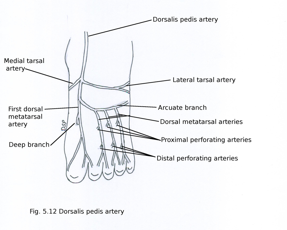
Dorsalis pedis artery
It is a continuation of anterior tibial artery crossing ankle joint goes up to proximal part of first intermetatarsal space. Here it goes to sole by passing in between two heads of first dorsal interosseous muscle. It completes plantar arch by joining with deep branch of lateral plantar artery.
Branches : 1) Lateral tarsal artery 2) Medial tarsal artery 3) Arcuate branch 4) First dorsal metatarsal artery 5) Small cutaneous branches
1) Lateral tarsal artery : Lateral tarsal artery goes laterally deep to extensor digitorum brevis and anastomoses with arcuate artery, lateral malleolar artery branch of anterior tibial artery, lateral plantar artery and perforating branches of peroneal artery to form lateral malleolar network.
2) Medial tarsal arteries : Two or three medial tarsal arteries goes towards medial side of ankle anastomoses with medial malleolar branch of anterior tibial artery, medial plantar artery, malleolar and calcaneal artery branches of posterior tibial artery to form medial malleolar network.
3) Arcuate branch : Arcuate artery arises near medial cuneiform bone and goes laterally over the bases of metatarsal bones. It lies deep to extensor tendons for digits. It anastomoses with lateral tarsal artery and branches of lateral plantar artery. It shows a convex course distally. It gives out second to fourth dorsal metatarsal arteries. After passing over dorsal interossei muscles in interdigital cleft it gives two dorsal digital arteries. Dorsal metatarsal arteries anastomoses with branches of plantar arch with the help of three proximal perforating arteries and with plantar metatarsal arteries with the help of four distal perforating arteries. Fourth dorsal metatarsal artery gives a small branch on lateral side of fifth toe.
4) First dorsal metatarsal artery : It is a branch from dorsalis pedis artery before its entry in sole. It goes distally over first dorsal interosseous muscle in first interdigital cleft between great toe and second toe here gives out dorsal digital branches for respective side of great toe and second toe. It also gives out a branch on medial side of great toe during its course.
5) Small cutaneous branches : Dorsalis pedis artery and its branch first dorsal metatarsal artery gives cutaneous branches to supply skin of dorsum of foot between extensor retinaculum and first interdigital cleft.
Terminal branches of Deep peroneal nerve
Deep peroneal nerve goes downwards in relation with lateral side of dorsalis pedis artery. It lies deep to extensor retinaculum. It divides into two branches lateral and medial terminal branches. Lateral terminal branch of deep peroneal nerve lies deep to extensor digitorum brevis and forms a pseudo ganglion then supply that muscle from ganglion three interosseous branches arises to supply tarsal and metatarsophalangeal joints of middle three toes. Medial terminal branch runs down medial to dorsalis pedis artery. Gives out two dorsal digital nerves to supply adjacent sides of great toe and second toe. It also gives out branches to first metatarsophalangeal joint and first dorsal interosseous muscle.
Deep peroneal nerve goes downwards in relation with lateral side of dorsalis pedis artery. It lies deep to extensor retinaculum. It divides into two branches lateral and medial terminal branches. Lateral terminal branch of deep peroneal nerve lies deep to extensor digitorum brevis and forms a pseudo ganglion then supply that muscle from ganglion three interosseous branches arises to supply tarsal and metatarsophalangeal joints of middle three toes. Medial terminal branch runs down medial to dorsalis pedis artery. Gives out two dorsal digital nerves to supply adjacent sides of great toe and second toe. It also gives out branches to first metatarsophalangeal joint and first dorsal interosseous muscle.
