LUNGS
There are two lungs right and left. These help in respiration. Right and left lung invaginated by pleura separately. These are elastic in nature and spongy in texture. In adult these are grayish in color because of deposition of carbon particles in it. Weight of right lung is 625 gm and left lung slightly less about 575 gm.
Parts of lung :
Each lung is conical in shape. Shows following parts 1) Apex 2) Base 3) Two surfaces costal and medial 4) Three borders Anterior, Posterior and Inferior
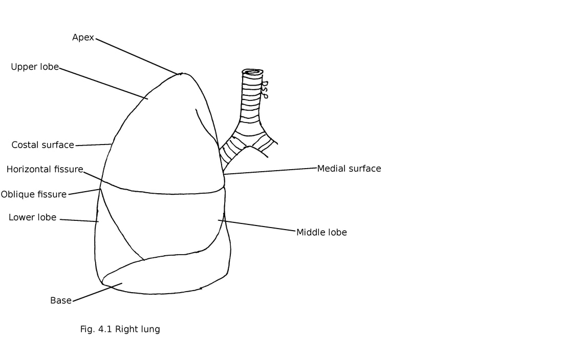
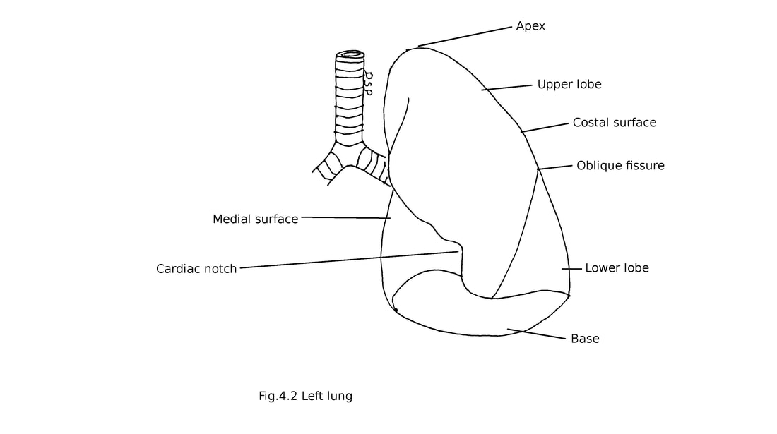
1) Apex : It is rounded upper part of lung. It lies about 3-4 cm above first costal cartilage. Apex lies in the root of neck. It is covered by cervical pleura supported by suprapleural membrane from outside. Subclavian artery goes upwards and laterally forming a groove.
2) Base : It is a concave shaped lower part of lung. Concavity is more on right side because liver present on right side. It is present over upper surface of diaphragm. Diaphragm separates base of right lung from right lobe of liver and base of left lung from left lobe of liver, fundus of stomach and spleen.
3) a) Costal surface : It is an outer surface of lung. It is convex and in contact with thoracic wall. It is covered by costal pleura. It shows transverse impressions for ribs.
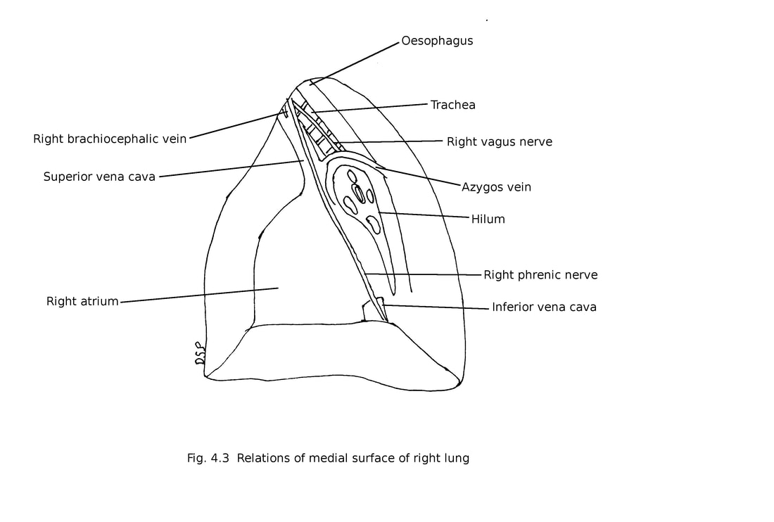
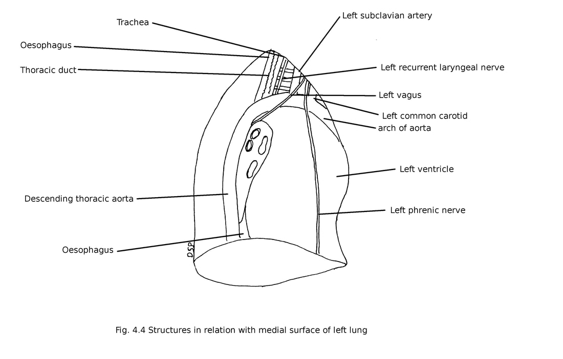
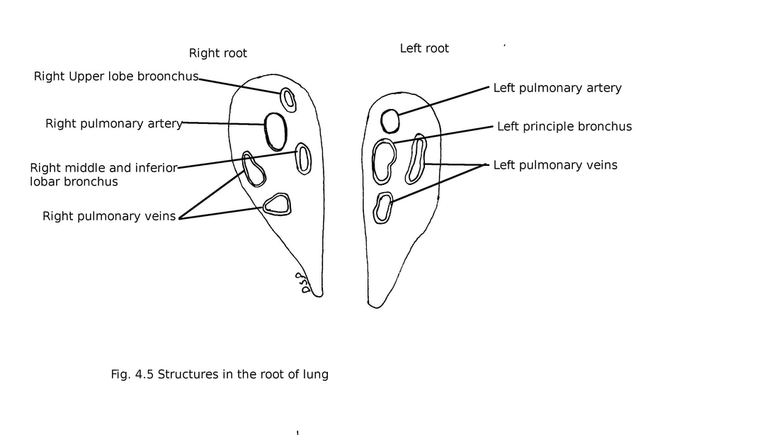
b) Medial surface : It shows posterior vertebral and anterior mediastinal part. Vertebral part comes in relation with sides of upper ten thoracic vertebra also inter-vertebral discs, posterior intercostal vessels and splanchnic nerves. Mediastinal part shows a deep concave impression for heart known as cardiac impression in left lung. Hilum is present on this part. Hilum is triangular in shape through which structures of lung root enter or exit. Following are structures present in right hilum of lung from above downwards upper lobe bronchus, pulmonary artery, middle and lower lobar bronchus and pulmonary vein. In right lung from before backwards upper pulmonary vein, pulmonary artery and bronchus. In left lung structures from above downwards pulmonary artery, left principal bronchus and lower pulmonary vein. In left lung from before backwards upper pulmonary vein, pulmonary artery and bronchus.
Structures in relation with medial surface of lung :
Following structures comes in relation with medial surface of right lung right atrium and right auricle, part of right ventricle, terminal part of inferior vena cava, right phrenic nerve, superior vena cava, lower part of right brachiocephalic vein, azygos vein, oesophagus, trachea and right vagus nerve.
2) Base : It is a concave shaped lower part of lung. Concavity is more on right side because liver present on right side. It is present over upper surface of diaphragm. Diaphragm separates base of right lung from right lobe of liver and base of left lung from left lobe of liver, fundus of stomach and spleen.
3) a) Costal surface : It is an outer surface of lung. It is convex and in contact with thoracic wall. It is covered by costal pleura. It shows transverse impressions for ribs.
b) Medial surface : It shows posterior vertebral and anterior mediastinal part. Vertebral part comes in relation with sides of upper ten thoracic vertebra also inter-vertebral discs, posterior intercostal vessels and splanchnic nerves. Mediastinal part shows a deep concave impression for heart known as cardiac impression in left lung. Hilum is present on this part. Hilum is triangular in shape through which structures of lung root enter or exit. Following are structures present in right hilum of lung from above downwards upper lobe bronchus, pulmonary artery, middle and lower lobar bronchus and pulmonary vein. In right lung from before backwards upper pulmonary vein, pulmonary artery and bronchus. In left lung structures from above downwards pulmonary artery, left principal bronchus and lower pulmonary vein. In left lung from before backwards upper pulmonary vein, pulmonary artery and bronchus.
Structures in relation with medial surface of lung :
Following structures comes in relation with medial surface of right lung right atrium and right auricle, part of right ventricle, terminal part of inferior vena cava, right phrenic nerve, superior vena cava, lower part of right brachiocephalic vein, azygos vein, oesophagus, trachea and right vagus nerve.
Following structures in relation with medial surface of left lung left ventricle, left auricle, part of right ventricle, pulmonary trunk, arch of aorta, descending thoracic aorta, left subclavian artery, thoracic duct, left brachiocephalic vein, left vagus nerve, oesophagus, left phrenic nerve, left recurrent laryngeal nerve.
4) Borders :
Anterior : Anterior border is thin. It is vertical on right side. But on left side it follows the line of reflection of pleura and show a cardiac notch. So it is a straight up to fourth costal cartilage then deviate laterally for 2.5 cm.
Posterior : It is thick and not so will defined. It separates costal surface from mediastinal surface. It comes in relation with heads of upper ten ribs.
Inferior : It is thin and sharp border. It separates base of lung from costal surface and medial surface. It follows a line which passes through 6th rib in midclavicular line, 8th rib in mid axillary line and a point 2 centimetre lateral to 10th thoracic spine.
Lobes and fissures of lungs
Right lung shows three lobes upper, middle and lower separated by two fissures oblique and horizontal. Left lung shows two lobes separated by one oblique fissure. Oblique fissure starts at the level of 4th thoracic vertebrae (below apex) in relation with posterior border. It separates lower lobe from upper and middle lobes. It runs downwards along with 5th intercostal space follow 6th rib to 6th costochondral junction on costal surface upto interior border 7.5 centimetre behind its anterior end. Horizontal fissure separates superior and middle lobes. It lies at the level of 4th costal cartilage starts from anterior border and meets with oblique fissure at mid axillary line. It is present only in right lung
Root of lung
It connects medial surface of lung to trachea and heart. It mainly contains principal bronchus, pulmonary artery, two pulmonary veins , bronchial vessels, pulmonary plexus, lymphatics, nerves and connective tissues. It lies in relation with bodies of 5th, 6th and 7th thoracic vertebra.
Structures present in root are already described in hilum of lung.
Structure of lungs
Each lung shows from outside inwards serous coat, sub-serous areolar tissue and pulmonary substance. Serious coat is formed by pulmonary pleura . Sub-serous tissues are formed by fibroelastic tissue. It gives fibroelastic septum inside lung substance forming small lung lobules.
Trachea divides into two principal bronchus at the level of fourth thoracic vertebra. Right principal bronchus is more wide and vertical. While left principal bronchus more oblique. Principal bronchus after passing through hilum divides into secondary bronchi or lobar bronchi 3 on right side and 2 on left side. Lobar bronchus divides into tertiary or segmental bronchi. There is one for each bronchopulmonary segment. Segmental bronchi then divided into smaller unit known as terminal bronchioles. The terminal bronchioles again divides and form respiratory bronchioles.
Arterial supply of lungs : Lung receives its arterial supply up to respiratory bronchioles from mainly bronchial arteries. On right side one bronchial artery which is a branch of third posterior intercostal artery or upper left bronchial artery. On left side two bronchial arteries which are branches coming from descending thoracic aorta. It supply bronchial tree and pulmonary tissue. It carries oxygenated blood for the nutrition of lung.
Respiratory part of lung supplied by pulmonary arteries. Pulmonary arteries carry deoxygenated blood and it drains into pulmonary capillary plexus.
Venous drainage of lungs :
Bronchial veins which drains deoxygenated blood to systemic veins with help of superficial and deep bronchial veins. Superficial bronchial veins receive blood from pleura and extra-pulmonary part of bronchi. On right side it drains into arch of azygos vein and on left side it drains into left superior intercostal vein or superior hemiazygos vein. Deep bronchial vein receive blood from intrapulmonary bronchi and bronchioles drains into pulmonary veins. Oxygenated blood from lungs drained by pulmonary veins.
Lymphatic drainage of lungs:
Superficial and deep group of lymph vessels drains bronchopulmonary group of lymph nodes. Superficial lymph vessels drains outer part of lung substance which lies in relation with pulmonary pleura. Deep lymph vessels drains from intrapulmonary bronchi, bronchioles and interlobular septum. Bronchopulmonary lymph node finally drains into superior and inferior tracheobronchial nodes.
Nerve supply of lungs :
Lung receives autonomic nerve supply. Sympathetic nerves supply comes from T2 to T5 spinal segments and parasympathetic nerve supply from vagus nerve. Parasympathetic fibres are motor in nature causes bronchoconstriction and secretion of bronchial glands. Sympathetic fibres causes bronchodilation and vasomotor in nature.
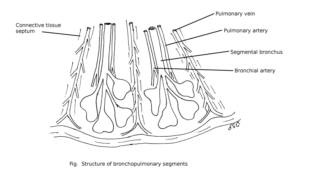
Bronchopulmonary segments
Bronchopulmonary segments are pyramidal shaped functional units of lungs. Its apex directed towards root of lung. It is mediated by terminal or segmental bronchi.
Structure of bronchopulmonary segment :
Pulmonary segments are pyramidal in shape divided by connective tissue septum. In this septum pulmonary veins are present. Each segment receives segmental bronchus, pulmonary vein and bronchial artery.
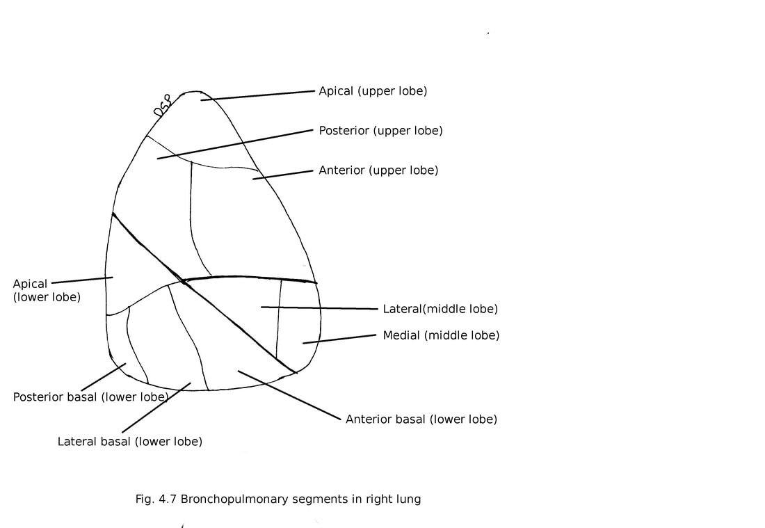
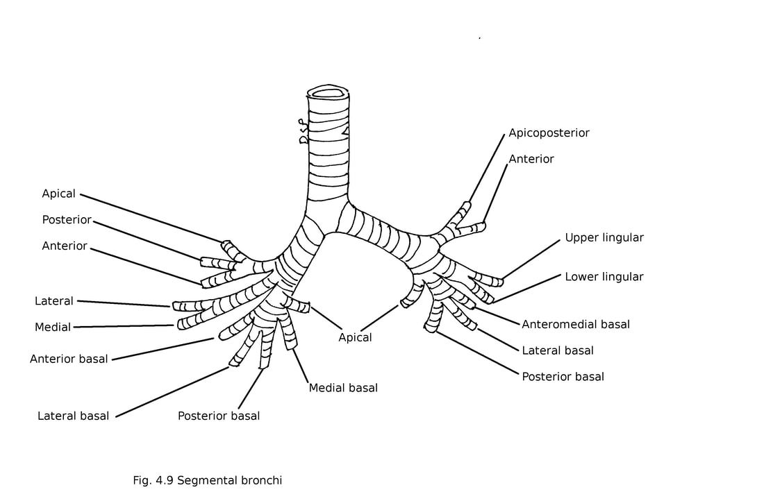
Bronchopulmonary segments of right lung :
These are 10 in number .
Upper lobe : 1) Apical, 2) Posterior, 3) Anterior
Middle lobe : 4) Lateral, 5) Medial
Lower lobe: 6) Apical, 7) Medial basal, 8) Lateral basal, 9) Posterior basal, 10) Anterior basal
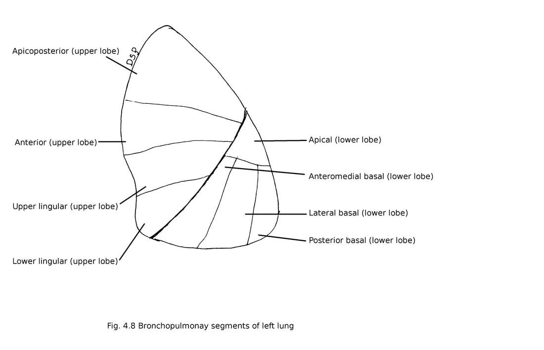
Bronchopulmonary segments of left lung :
These are 8 (may be 10) in number .
Upper lobe : 1) Apicoposterior, 2) Anterior, 3) Upper lingular, 4) Lower lingular
Lower lobe : 5) Apical, 6) Anteromedial basal, 7) Lateral basal, 8) Posterior basal
Bronchopulmonary segments are pyramidal shaped functional units of lungs. Its apex directed towards root of lung. It is mediated by terminal or segmental bronchi.
Structure of bronchopulmonary segment :
Pulmonary segments are pyramidal in shape divided by connective tissue septum. In this septum pulmonary veins are present. Each segment receives segmental bronchus, pulmonary vein and bronchial artery.
Bronchopulmonary segments of right lung :
These are 10 in number .
Upper lobe : 1) Apical, 2) Posterior, 3) Anterior
Middle lobe : 4) Lateral, 5) Medial
Lower lobe: 6) Apical, 7) Medial basal, 8) Lateral basal, 9) Posterior basal, 10) Anterior basal
Bronchopulmonary segments of left lung :
These are 8 (may be 10) in number .
Upper lobe : 1) Apicoposterior, 2) Anterior, 3) Upper lingular, 4) Lower lingular
Lower lobe : 5) Apical, 6) Anteromedial basal, 7) Lateral basal, 8) Posterior basal
Applied anatomy :
- Lung abscess commonly occurs in apical segment of right lower lobe and posterior segment of right upper lobe.
- Tuberculosis affects posterior segment of right upper lobe.
- Bronchopulmonary segments acts as a barrier to the spread of infection from one segment to another but tuberculosis is exception.
- For postural drainage of abscess and surgical removal of segment knowledge of bronchopulmonary segments in essential.
