MAJOR BLOOD VESSELS
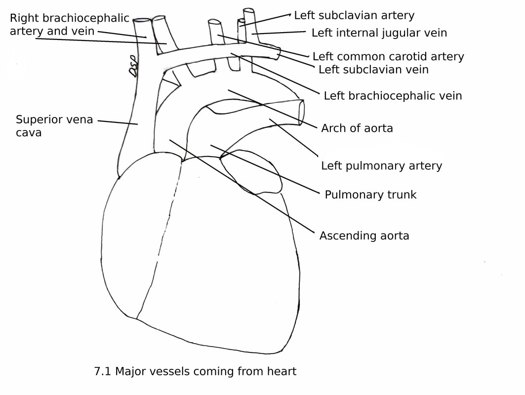
Superior vena cava
It is a large vein draining venous blood from the upper part of the body into the right atrium. It is formed behind the lower border of the first right costal cartilage near sternum by joining right and left brachiocephalic veins. It is about 7 cm in length. It pierces the fibrous pericardium near the right second costal cartilage.
Relations :
Anterior : Right Internal thoracic vessels, Anterior margin of the right lung and Pleura, part of pericardium in lower aspect.
Posterior : Trachea, right vagus nerve, root of right lung in lower part
Medial (Left) : Brachiocephalic trunk, Ascending aorta.
Lateral (Right) : Phrenic nerve with pericardiophrenic vessels, right lung and right pleura.
Tributaries :
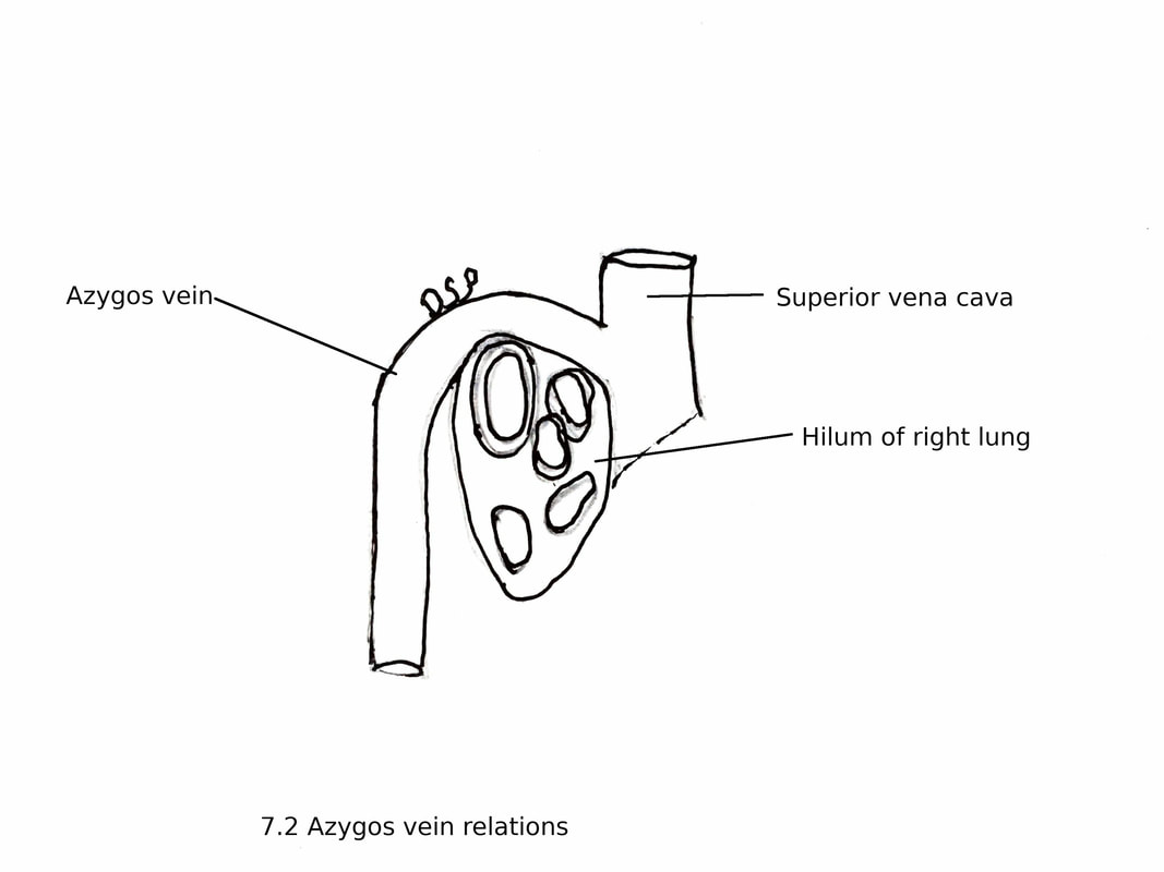
I. The azygos vein : Opens into the superior vena cava at second costal cartilage after arching over the root of the right lung.
2. Right and left brachio-cephalic veins
3. Small Pericardial veins and Mediastinal veins.
Applied Anatomy
1. Obstruction : When obstruction is above the opening of the azygos vein. Collateral circulation established through azygos vein via internal thoracic vein, anterior intercostal veins, posterior intercostal veins then azygos vein. So connecting the upper part of the body.
When obstruction is below the opening of the azygos vein collateral circulation established through azygos vein to inferior vena cava via superior and inferior epigastric veins, lateral thoracic veins, thoraco epigastric veins.
Facial edema and facial congestion is common in it. Common cause is bronchial carcinoma.
It is a large vein draining venous blood from the upper part of the body into the right atrium. It is formed behind the lower border of the first right costal cartilage near sternum by joining right and left brachiocephalic veins. It is about 7 cm in length. It pierces the fibrous pericardium near the right second costal cartilage.
Relations :
Anterior : Right Internal thoracic vessels, Anterior margin of the right lung and Pleura, part of pericardium in lower aspect.
Posterior : Trachea, right vagus nerve, root of right lung in lower part
Medial (Left) : Brachiocephalic trunk, Ascending aorta.
Lateral (Right) : Phrenic nerve with pericardiophrenic vessels, right lung and right pleura.
Tributaries :
I. The azygos vein : Opens into the superior vena cava at second costal cartilage after arching over the root of the right lung.
2. Right and left brachio-cephalic veins
3. Small Pericardial veins and Mediastinal veins.
Applied Anatomy
1. Obstruction : When obstruction is above the opening of the azygos vein. Collateral circulation established through azygos vein via internal thoracic vein, anterior intercostal veins, posterior intercostal veins then azygos vein. So connecting the upper part of the body.
When obstruction is below the opening of the azygos vein collateral circulation established through azygos vein to inferior vena cava via superior and inferior epigastric veins, lateral thoracic veins, thoraco epigastric veins.
Facial edema and facial congestion is common in it. Common cause is bronchial carcinoma.
Thoracic part of aorta
The aorta is a big arterial trunk carrying oxygenated blood from the left ventricle. It shows three parts a) Ascending aorta, b) Arch of the aorta and c) Descending aorta.
Ascending aorta starts from the left ventricle. It is about 5cm in length. It starts posterior to the left half of the sternum at lower border of the third costal cartilage. Then it goes upwards, forwards and towards right behind the left half of sternum upto upper border of the right second costal cartilage. Then it continues as an arch of the aorta.
Near the root of the ascending aorta there are three aortic sinuses present. Aortic sinuses are three dilatations in relation with an aortic wall. These are anterior, left posterior and right posterior.
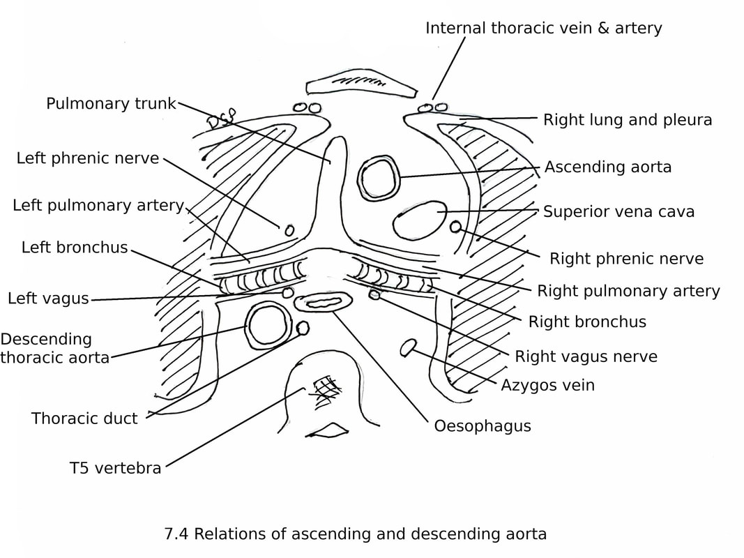
Relations :
Anterior : Sternum, left lung and pleura, infundibulum of the right ventricle, right auricle and root of the pulmonary trunk.
Posterior : Right principal bronchus, transverse sinus, left atrium and right pulmonary artery
Right side : Right atrium and superior vena cava,
Left side : Left atrium and pulmonary trunk
Branches :
1. Right coronary artery (branch of the anterior aortic sinus)
2. Left coronary artery ( branch of the left posterior aortic sinus)
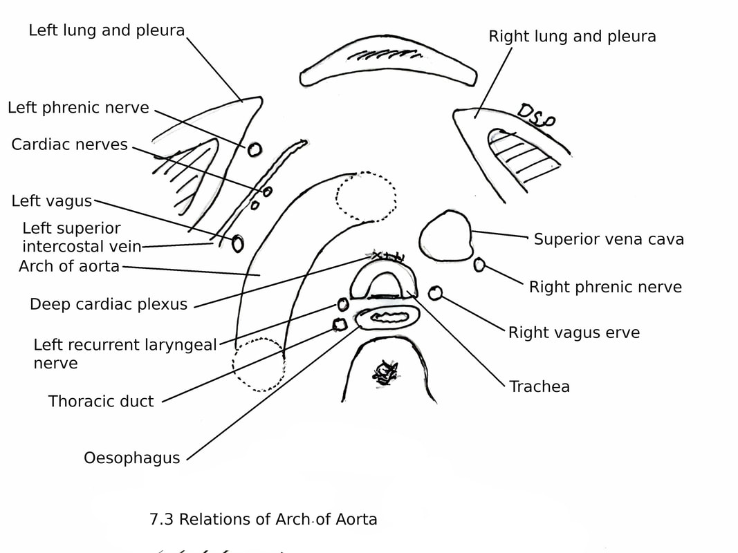
Arch of the Aorta
Ascending aorta continues as an arch of aorta by arching over the root of the left lung. It lies in the superior mediastinum posterior to the manubrium sterni.
It starts posterior to the right second sternochondral joint. Then it runs upwards, backwards and to the left. Here it lies anterior to the trachea then goes to the left side of the bifurcation of the trachea. After passing downwards behind the left bronchus it arches over the root of the left lung at a lower margin of the body of the 4th thoracic vertebra. It continues as a descending aorta.
Relations :
Anteriorly and to the left:
1. Crossed by four nerves and one vein. These are left phrenic, Inferior cervical cardiac branch of the left vagus, superior cervical cardiac branch of left sympathetic chain, left vagus and left superior intercostal vein.
2. Left pleura and lung.
3. Remains of thymus.
Posteriorly and to the right :
1. Trachea close to bifurcation
2. Deep cardiac plexus
3. Oesophagus.
4. Left recurrent laryngeal nerve.
5. Thoracic duct.
6. Fourth thoracic vertebra
Superior :
1. Origin of brachiocephalic trunk, left common carotid and left subclavian arteries.
2. Arteries are crossed in front by the left brachiocephalic vein.
3. Crossing of the left phrenic and left vagus.
Inferior :
1. Bifurcation of the pulmonary trunk.
2. Left principal bronchus.
3. Ligamentum arteriosum
4. Superficial cardiac plexus
5. Left recurrent laryngeal nerve.
Branches :
1. Brachiocephalic trunk with its two branches right common carotid artery and right subclavian artery
2. Left common carotid artery.
3. Left subclavian artery.
4. Occasional thyroidea ima artery, vertebral artery, internal thoracic artery may arise from it.
Descending Thoracic Aorta
Arch of the aorta continues as descending thoracic aorta. It is present in the posterior mediastinum.
At the level of the lower border of the body of the 4th thoracic vertebra on its left side it starts. It goes downwards and terminates at the lower border of the 12th thoracic vertebra after passing through diaphragmatic aortic aperture.
Relations :
Anterior : Pericardium, root of left lung, oesophagus and diaphragm.
Posterior : Hemiazygos veins and vertebral column
Right side: Right lung and pleura, oesophagus, azygos vein and thoracic duct.
Left side: Left lung and pleura.
Branches :
1. Pericardial branches to pericardium
2. Bronchial arteries : Left bronchial arteries two in number arises from thoracic aorta.
3. Posterior intercostal arteries nine pairs in 3rd to 11th intercostal spaces.
4. Two subcostal arteries : These are the last paired branches. Enter the abdomen below the lower border of 12th rib with 12th thoracic nerve deep to lateral arcuate ligament.
5. Oesophageal branches
6. Mediastinal branches which will supply lymph nodes and areolar tissue in posterior mediastinum.
7. Phrenic arteries : Which will supply the upper surface of the diaphragm. Also gives out branches to anastomose with musculophrenic and pericardiophrenic
arteries.
Applied Anatomy :
1. Aortic knuckle: A convex shadow is visible in PA view of X-ray chest on left side of sternal angle known as aortic knuckle. It is formed by the arch of aorta.
2. Aneurysm of aorta is a localized abnormal dilatation. It may cause pressure on surrounding structures. Cause may be abnormalities of connective tissues.
3. Coarctation of the aorta is localized constriction of aorta. It may be near or distal to attachment of the ductus arteriosus. In preductal type patent ductus arteriosus present.
4. Aortic dissection : In it dissection of aorta occurs due to degeneration of inner wall of aorta. There occur splitting of wall and creating false lumen.
5. Aortic calcification : Atherosclerosis formed plaque gets calcified. Common in descending aorta.
Pulmonary trunk
The pulmonary trunk, or pulmonary artery, carries deoxygenated blood from the right ventricle to the right and left lungs. It is short and wide approximately 5 centimetres in length and 3 centimetres in diameter. It originates from the right ventricle. Then it goes upward and backward in relation with ascending aorta in front then on its left. Just below the aortic arch it divides into right and left pulmonary arteries. Here it lies in frontolateral aspect of tracheal bifurcation.
Pulmonary arteries
There are two pulmonary arteries right and left. The right pulmonary artery shows two branches superior and inferior. It lies posterior to superior vena cava. Superior artery supply upper lobe forming major supply of that lobe. The inferior artery gives a branch to the middle lobe and a branch to the lower lobe. Inferior branch also gives a small recurrent branch to the upper lobe.
The left pulmonary artery lies anterior to the descending aorta. It gives two or more branches. A large branch to the left upper lobe, lingular branches and separate branches supply lower lobe.
The aorta is a big arterial trunk carrying oxygenated blood from the left ventricle. It shows three parts a) Ascending aorta, b) Arch of the aorta and c) Descending aorta.
Ascending aorta starts from the left ventricle. It is about 5cm in length. It starts posterior to the left half of the sternum at lower border of the third costal cartilage. Then it goes upwards, forwards and towards right behind the left half of sternum upto upper border of the right second costal cartilage. Then it continues as an arch of the aorta.
Near the root of the ascending aorta there are three aortic sinuses present. Aortic sinuses are three dilatations in relation with an aortic wall. These are anterior, left posterior and right posterior.
Relations :
Anterior : Sternum, left lung and pleura, infundibulum of the right ventricle, right auricle and root of the pulmonary trunk.
Posterior : Right principal bronchus, transverse sinus, left atrium and right pulmonary artery
Right side : Right atrium and superior vena cava,
Left side : Left atrium and pulmonary trunk
Branches :
1. Right coronary artery (branch of the anterior aortic sinus)
2. Left coronary artery ( branch of the left posterior aortic sinus)
Arch of the Aorta
Ascending aorta continues as an arch of aorta by arching over the root of the left lung. It lies in the superior mediastinum posterior to the manubrium sterni.
It starts posterior to the right second sternochondral joint. Then it runs upwards, backwards and to the left. Here it lies anterior to the trachea then goes to the left side of the bifurcation of the trachea. After passing downwards behind the left bronchus it arches over the root of the left lung at a lower margin of the body of the 4th thoracic vertebra. It continues as a descending aorta.
Relations :
Anteriorly and to the left:
1. Crossed by four nerves and one vein. These are left phrenic, Inferior cervical cardiac branch of the left vagus, superior cervical cardiac branch of left sympathetic chain, left vagus and left superior intercostal vein.
2. Left pleura and lung.
3. Remains of thymus.
Posteriorly and to the right :
1. Trachea close to bifurcation
2. Deep cardiac plexus
3. Oesophagus.
4. Left recurrent laryngeal nerve.
5. Thoracic duct.
6. Fourth thoracic vertebra
Superior :
1. Origin of brachiocephalic trunk, left common carotid and left subclavian arteries.
2. Arteries are crossed in front by the left brachiocephalic vein.
3. Crossing of the left phrenic and left vagus.
Inferior :
1. Bifurcation of the pulmonary trunk.
2. Left principal bronchus.
3. Ligamentum arteriosum
4. Superficial cardiac plexus
5. Left recurrent laryngeal nerve.
Branches :
1. Brachiocephalic trunk with its two branches right common carotid artery and right subclavian artery
2. Left common carotid artery.
3. Left subclavian artery.
4. Occasional thyroidea ima artery, vertebral artery, internal thoracic artery may arise from it.
Descending Thoracic Aorta
Arch of the aorta continues as descending thoracic aorta. It is present in the posterior mediastinum.
At the level of the lower border of the body of the 4th thoracic vertebra on its left side it starts. It goes downwards and terminates at the lower border of the 12th thoracic vertebra after passing through diaphragmatic aortic aperture.
Relations :
Anterior : Pericardium, root of left lung, oesophagus and diaphragm.
Posterior : Hemiazygos veins and vertebral column
Right side: Right lung and pleura, oesophagus, azygos vein and thoracic duct.
Left side: Left lung and pleura.
Branches :
1. Pericardial branches to pericardium
2. Bronchial arteries : Left bronchial arteries two in number arises from thoracic aorta.
3. Posterior intercostal arteries nine pairs in 3rd to 11th intercostal spaces.
4. Two subcostal arteries : These are the last paired branches. Enter the abdomen below the lower border of 12th rib with 12th thoracic nerve deep to lateral arcuate ligament.
5. Oesophageal branches
6. Mediastinal branches which will supply lymph nodes and areolar tissue in posterior mediastinum.
7. Phrenic arteries : Which will supply the upper surface of the diaphragm. Also gives out branches to anastomose with musculophrenic and pericardiophrenic
arteries.
Applied Anatomy :
1. Aortic knuckle: A convex shadow is visible in PA view of X-ray chest on left side of sternal angle known as aortic knuckle. It is formed by the arch of aorta.
2. Aneurysm of aorta is a localized abnormal dilatation. It may cause pressure on surrounding structures. Cause may be abnormalities of connective tissues.
3. Coarctation of the aorta is localized constriction of aorta. It may be near or distal to attachment of the ductus arteriosus. In preductal type patent ductus arteriosus present.
4. Aortic dissection : In it dissection of aorta occurs due to degeneration of inner wall of aorta. There occur splitting of wall and creating false lumen.
5. Aortic calcification : Atherosclerosis formed plaque gets calcified. Common in descending aorta.
Pulmonary trunk
The pulmonary trunk, or pulmonary artery, carries deoxygenated blood from the right ventricle to the right and left lungs. It is short and wide approximately 5 centimetres in length and 3 centimetres in diameter. It originates from the right ventricle. Then it goes upward and backward in relation with ascending aorta in front then on its left. Just below the aortic arch it divides into right and left pulmonary arteries. Here it lies in frontolateral aspect of tracheal bifurcation.
Pulmonary arteries
There are two pulmonary arteries right and left. The right pulmonary artery shows two branches superior and inferior. It lies posterior to superior vena cava. Superior artery supply upper lobe forming major supply of that lobe. The inferior artery gives a branch to the middle lobe and a branch to the lower lobe. Inferior branch also gives a small recurrent branch to the upper lobe.
The left pulmonary artery lies anterior to the descending aorta. It gives two or more branches. A large branch to the left upper lobe, lingular branches and separate branches supply lower lobe.
