MEDIASTINUM
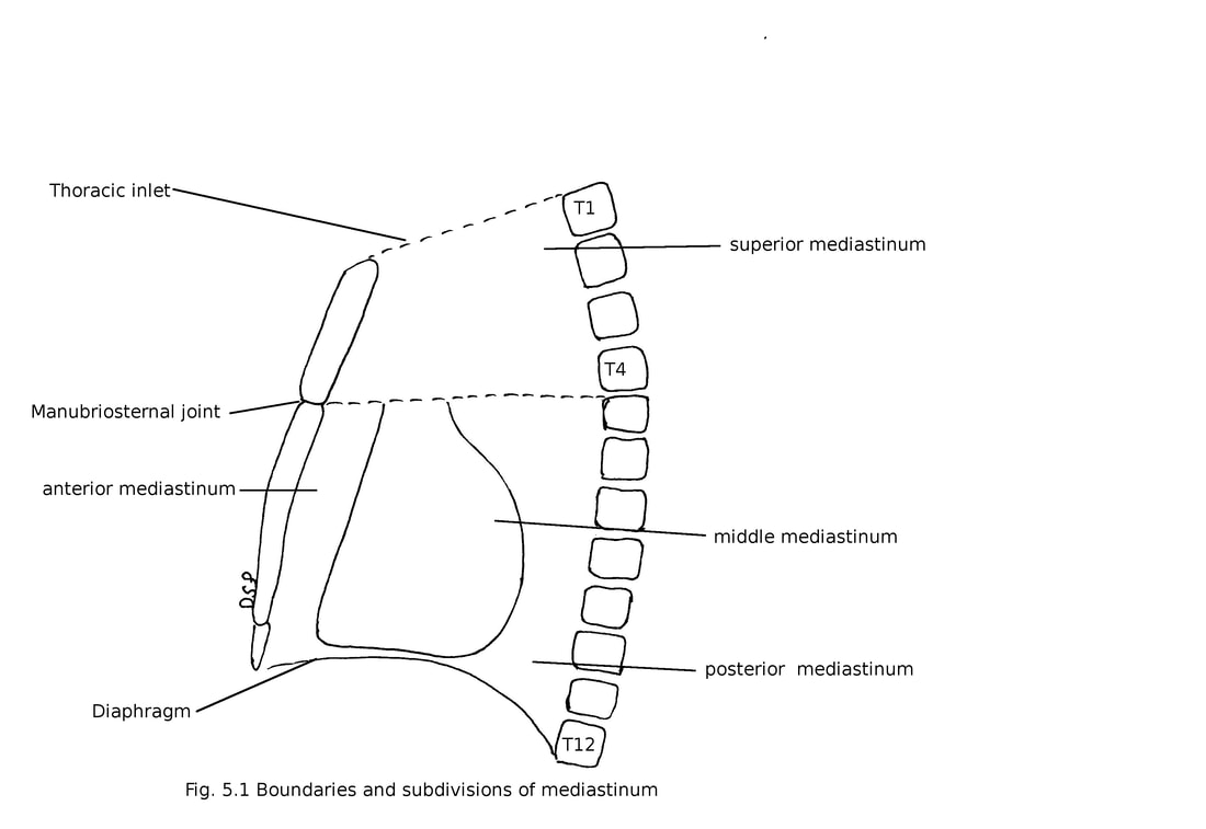
Mediastinum is a region between right pleural sac and left pleural sac. On each side covered by mediastinal pleura. Sternum lies anteriorly and twelve thoracic vertebrae posteriorly. It extends from thoracic inlet above up to diaphragm below.
Boundaries:
Anteriorly : Sternum
Posteriorly : Twelve thoracic vertebra
Above : Thoracic inlet
Below : Diaphragm
Both side : Mediastinal pleura
Mediastinum is divided into superior and inferior mediastinum by a plane which passes through manubriosternal joint and lower border of fourth thoracic vertebra. Inferior mediastinum again divided into 3 parts anterior, middle and posterior by pericardium.
Superior mediastinum :
Boundaries:
Anteriorly : Manubrium Sternum
Posteriorly : Upper four thoracic vertebra
Above : Thoracic inlet
Below : Plane which passes through manubriosternal joint to lower border of fourth thoracic vertebra
Both side : Mediastinal pleura
Contents
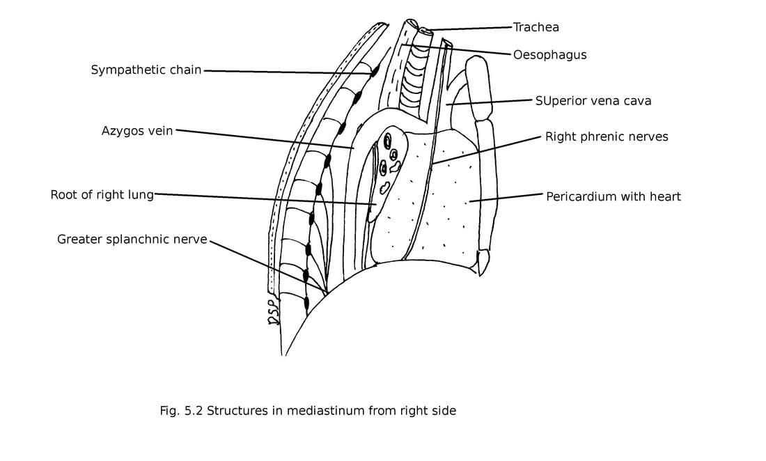
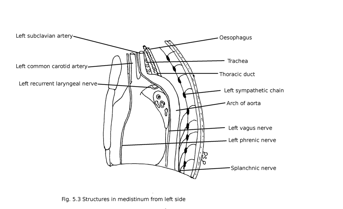
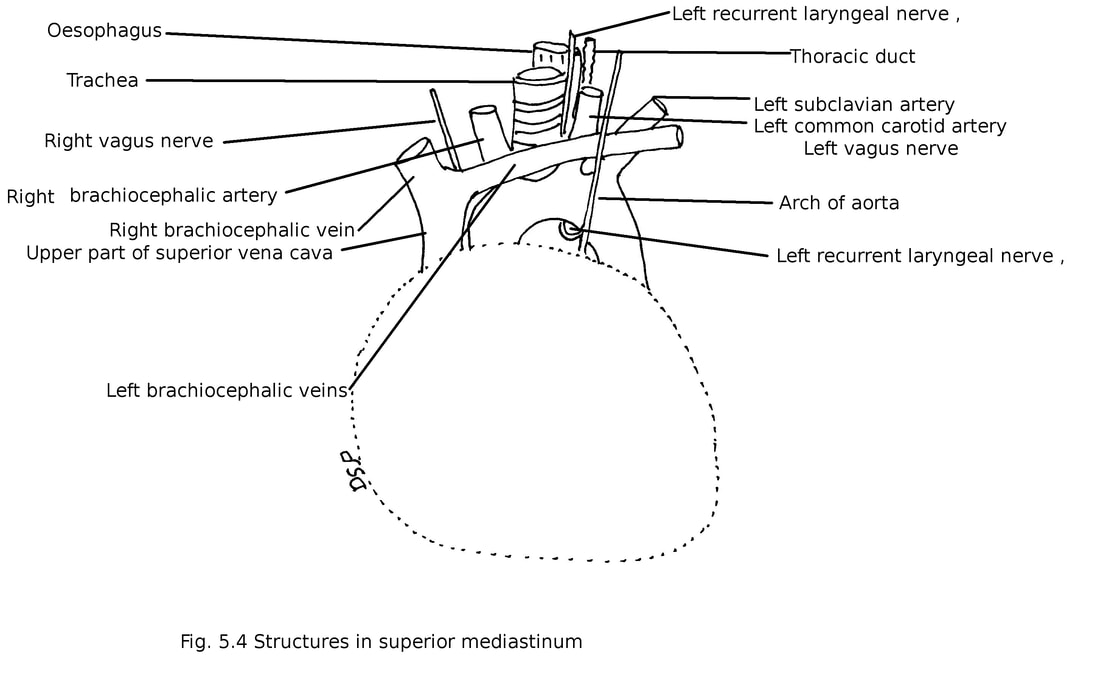
Trachea
Oesophagus
Thymus gland
Thoracic duct
Origin of sternohyoid and sternothyroid muscles, longus colli muscle
Nerves : Left recurrent laryngeal nerve , vagus nerve, phrenic nerve, cardiac nerves, cardiac plexus
Arteries : Arch of aorta, brachiocephalic artery, left subclavian artery, common carotid artery, internal thoracic arteries.
Veins : Right and left brachiocephalic veins, upper part of superior vena cava, left superior intercostal vein, internal thoracic veins
Paratracheal and tracheobronchial lymph nodes.
Boundaries:
Anteriorly : Manubrium Sternum
Posteriorly : Upper four thoracic vertebra
Above : Thoracic inlet
Below : Plane which passes through manubriosternal joint to lower border of fourth thoracic vertebra
Both side : Mediastinal pleura
Contents
Trachea
Oesophagus
Thymus gland
Thoracic duct
Origin of sternohyoid and sternothyroid muscles, longus colli muscle
Nerves : Left recurrent laryngeal nerve , vagus nerve, phrenic nerve, cardiac nerves, cardiac plexus
Arteries : Arch of aorta, brachiocephalic artery, left subclavian artery, common carotid artery, internal thoracic arteries.
Veins : Right and left brachiocephalic veins, upper part of superior vena cava, left superior intercostal vein, internal thoracic veins
Paratracheal and tracheobronchial lymph nodes.
Inferior mediastinum
Anterior mediastinal
It is an a narrow space which lies in between body of sternum and fibrous pericardium. It is narrow above at the level of fourth costal cartilage and broad below because left pleural sac deviates laterally.
Boundaries :
Anteriorly: Body of Sternum
Posteriorly : Fibrous pericardium
Superiorly : An imaginary plane with divides superior mediastinum and inferior mediastinum Inferiorly : Upper surface of diaphragm
On each side : Mediastinal pleura
Contents :
Superior and inferior sternopericardial ligaments, loose connective tissue, lymph nodes, mediastinal branches of internal thoracic artery, lower part of thymus.
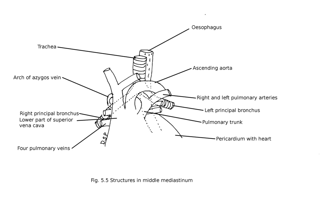
Middle mediastinum
It is a large part of mediastinum mainly occupied by pericardium, heart and other contents.
Contents :
Pericardium with heart
Arteries : Ascending aorta, pulmonary trunk, right and left pulmonary arteries.
Veins : Four pulmonary veins, lower part of superior vena cava, arch of azygos vein.
Nerves : Phrenic nerves, deep cardiac plexus.
Bifurcation of trachea, right and left principal bronchi, tracheobronchial lymph nodes.
Anterior mediastinal
It is an a narrow space which lies in between body of sternum and fibrous pericardium. It is narrow above at the level of fourth costal cartilage and broad below because left pleural sac deviates laterally.
Boundaries :
Anteriorly: Body of Sternum
Posteriorly : Fibrous pericardium
Superiorly : An imaginary plane with divides superior mediastinum and inferior mediastinum Inferiorly : Upper surface of diaphragm
On each side : Mediastinal pleura
Contents :
Superior and inferior sternopericardial ligaments, loose connective tissue, lymph nodes, mediastinal branches of internal thoracic artery, lower part of thymus.
Middle mediastinum
It is a large part of mediastinum mainly occupied by pericardium, heart and other contents.
Contents :
Pericardium with heart
Arteries : Ascending aorta, pulmonary trunk, right and left pulmonary arteries.
Veins : Four pulmonary veins, lower part of superior vena cava, arch of azygos vein.
Nerves : Phrenic nerves, deep cardiac plexus.
Bifurcation of trachea, right and left principal bronchi, tracheobronchial lymph nodes.
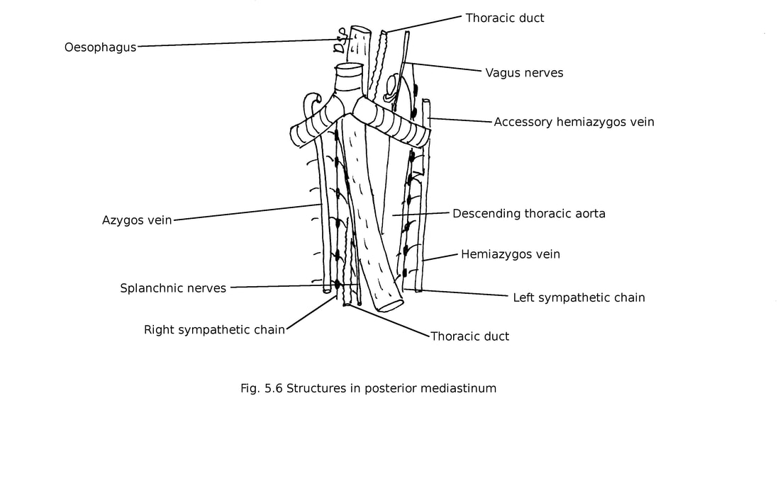
Posterior mediastinum
Boundaries :
Anteriorly : Fibrous pericardium, bifurcation of trachea, pulmonary vessels, posterior part of sloping upper surface of diaphragm.
Posteriorly : Lower eight thoracic vertebrae, anterior longitudinal ligament
On each side : Mediastinal pleura
Superiorly : An Imaginary plane with divides superior mediastinum and inferior mediastinum Inferiorly : Upper surface of diaphragm.
Contents :
Oesophagus
Arteries : Descending thoracic aorta with its branches.
Veins : Azygos vein, hemiazygos vein, accessory hemiazygos vein.
Nerves : Vagus nerves, splanchnic nerves, right and left sympathetic chains.
Posterior mediastinal group of lymph nodes, thoracic duct.
Applied anatomy :
1. Mediastinal shift : Mediastinum can shift towards diseased lung in disease like collapse of lung. It may shift towards opposite side in disease of pleura like pneumothorax or hydrothorax.
2. Infection can spread from neck to superior and posterior mediastinum because area in relation with vessels of neck, trachea, oesophagus are in communication with superior and posterior mediastinum.
3. Mediastinal syndrome : Compression of structures of mediastinum by tumour called mediastinal syndrome. Engorgement of veins of upper part of body, dyspnoea, cough, dysphagia, hoarseness of voice, paralysis of diaphragm, intercostal neuralgia. These symptoms are due to pressure on superior vena cava, trachea, oesophagus, left recurrent laryngeal nerve, phrenic nerve, intercostal nerves.
