PECTORAL REGION
It is anterior part of the chest cavity. Superficial fascia of the pectoral region is fibrofatty it contains mainly breast or mammary gland. The deep fascia of pectoral region also known as pectoral fascia it shows attachment on clavicle, sternum. It enclose pectoralis major muscle and also covers deltoid muscle. It also forms axillary fascia in axillary region and then it split to enclose latissimus dorsi muscle.
It is anterior part of the chest cavity. Superficial fascia of the pectoral region is fibrofatty it contains mainly breast or mammary gland. The deep fascia of pectoral region also known as pectoral fascia it shows attachment on clavicle, sternum. It enclose pectoralis major muscle and also covers deltoid muscle. It also forms axillary fascia in axillary region and then it split to enclose latissimus dorsi muscle.
Breast or mammary gland
Breast present on both side of pectoral region in male or female but in male it remains rudimentary. In female after puberty these becomes well developed and during pregnancy and lactation it increases in size. Brest lies in superficial fascia of pectoral region and it is a modified sweat gland. Extent circular base of the breast extends from 2nd to 6th rib in mid clavicular line horizontally from lateral border of sternum to mid axillary line of 4th rib.
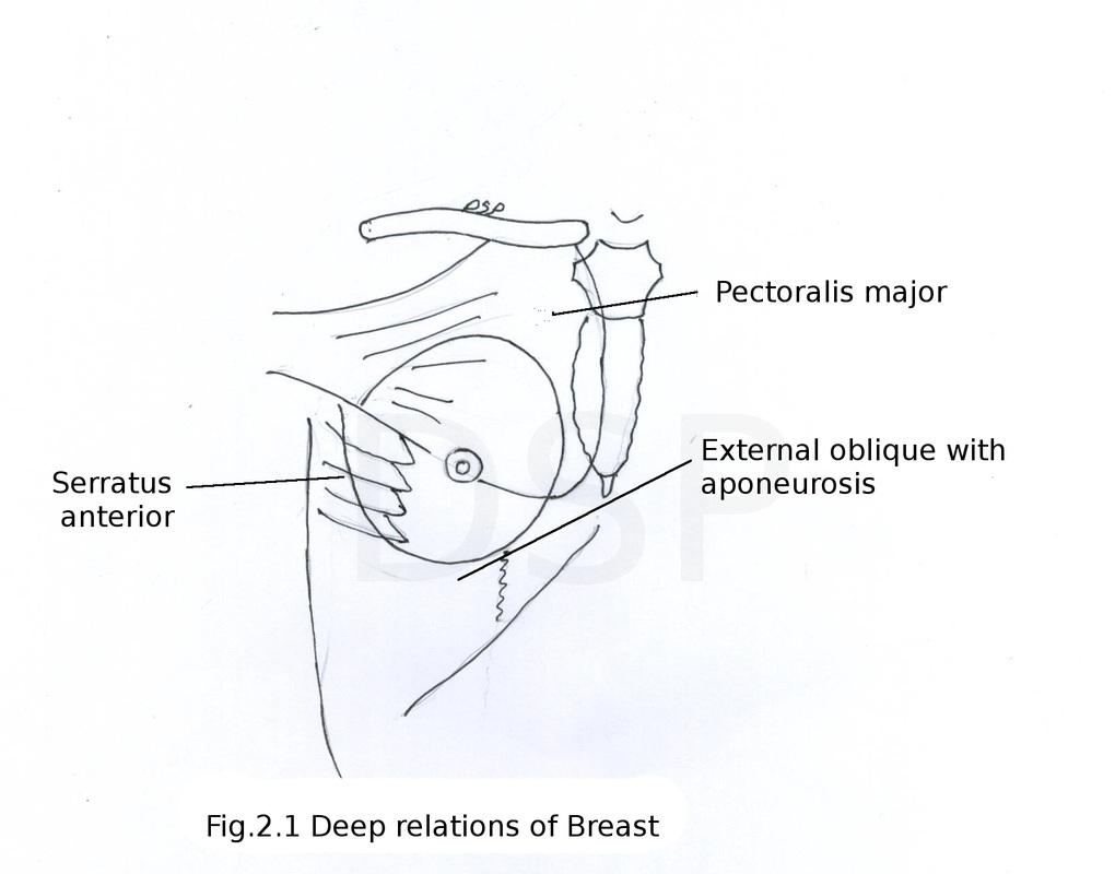
Deep structures part of the muscles lied deep to mammary gland serratus anterior, external oblique and pectoralis major.
There is a space in between mammary gland and deep fascia covering pectoralis major muscle so that breast can move over deep fascia covering pectoralis major. A part of breast extends into axilla called axillary tail of spence.
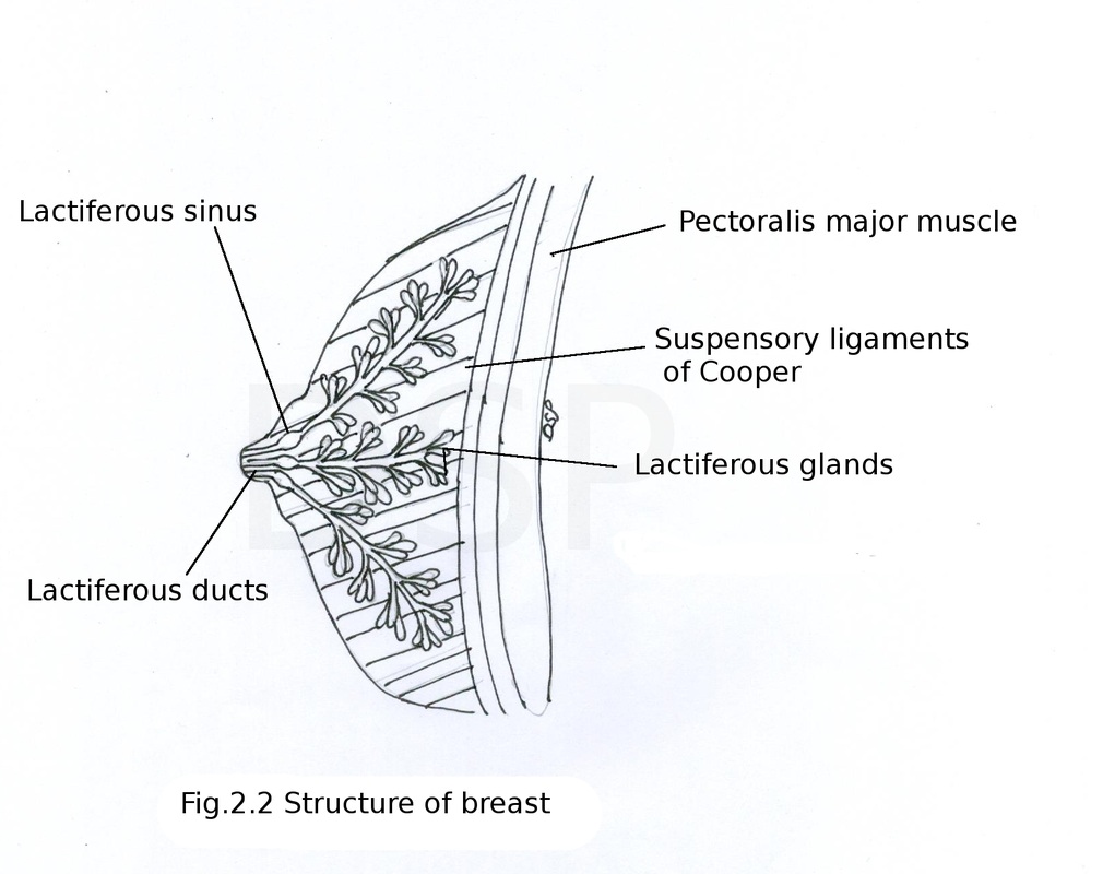
Structure of the breast
Skin of the breast shows nipple and areola. Nipple is the conical projection just below the centre of breast at the level of 4th intercostal space it contains 15 to 20 lactiferous ducts also circular and longitudinal smooth muscle fibres these muscle fibres elevate and depresses nipple respectively. It has rich nerve supply.
Areola it is circular dark pigmented area around nipple. It contains modified sebaceous glands these glands enlarge during pregnancy and lactation forming tubercles of montgomery. Skin of areola do not contains hair and fat.
Remaining part of breast formed by fatty tissue, fibrous tissue and glandular tissue. Fatty tissue forms main part of the mammary glands except areola and nipple. Fibrous tissue divide breast into different lobes by forming septa within substance of mammary gland it connects skin of breast and pectoral fascia known as suspensory ligament of cooper. Glandular tissue of breast form by tubuloalveolar type of glands. These glands are present in 15 to 20 lobes of breast draining in to lactiferous ducts. These ducts converge towards the nipple it shows a small dilatation called lactiferous sinus. This ducts finally open on to nipple.
Breast present on both side of pectoral region in male or female but in male it remains rudimentary. In female after puberty these becomes well developed and during pregnancy and lactation it increases in size. Brest lies in superficial fascia of pectoral region and it is a modified sweat gland. Extent circular base of the breast extends from 2nd to 6th rib in mid clavicular line horizontally from lateral border of sternum to mid axillary line of 4th rib.
Deep structures part of the muscles lied deep to mammary gland serratus anterior, external oblique and pectoralis major.
There is a space in between mammary gland and deep fascia covering pectoralis major muscle so that breast can move over deep fascia covering pectoralis major. A part of breast extends into axilla called axillary tail of spence.
Structure of the breast
Skin of the breast shows nipple and areola. Nipple is the conical projection just below the centre of breast at the level of 4th intercostal space it contains 15 to 20 lactiferous ducts also circular and longitudinal smooth muscle fibres these muscle fibres elevate and depresses nipple respectively. It has rich nerve supply.
Areola it is circular dark pigmented area around nipple. It contains modified sebaceous glands these glands enlarge during pregnancy and lactation forming tubercles of montgomery. Skin of areola do not contains hair and fat.
Remaining part of breast formed by fatty tissue, fibrous tissue and glandular tissue. Fatty tissue forms main part of the mammary glands except areola and nipple. Fibrous tissue divide breast into different lobes by forming septa within substance of mammary gland it connects skin of breast and pectoral fascia known as suspensory ligament of cooper. Glandular tissue of breast form by tubuloalveolar type of glands. These glands are present in 15 to 20 lobes of breast draining in to lactiferous ducts. These ducts converge towards the nipple it shows a small dilatation called lactiferous sinus. This ducts finally open on to nipple.
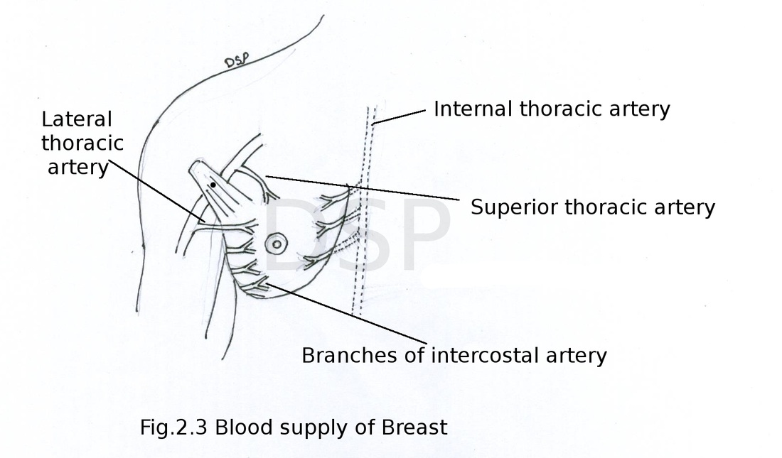
Arterial supply of breast:
superior thoracic artery, acromiothoracic artery and lateral thoracic artery branches of axillary artery. Perforating branches of internal thoracic artery.
Venous drainage:
Veins form plexus in relation to areola these forms superficial veins and deep veins drains into internal thoracic vein, intercostal vein, and axillary veins.
Nerve supply:
Nerves are coming from anterior and lateral cutaneous branches of 4th to 6th intercostal nerves. These carry sensory fibres to the skin and autonomic fibres which are vasomotor to muscles and blood vessels.
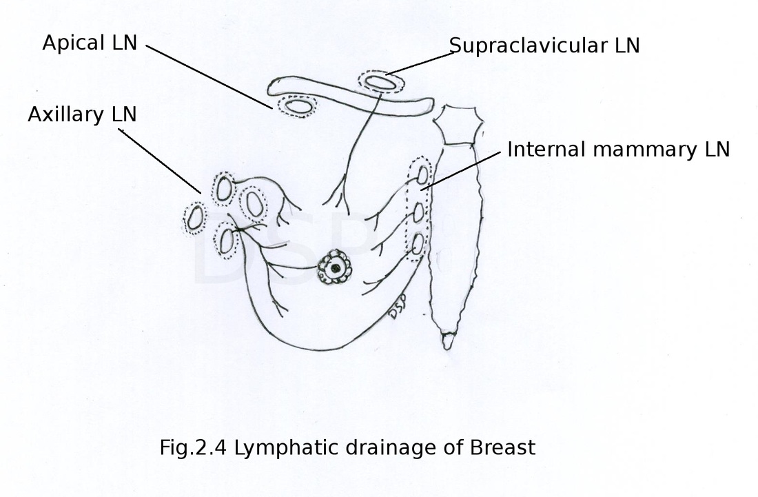
Lymphatic drainage:
Two sets of lymphatic drainage breast skin and substance of breast.From skin except areola and nipple. Lateral part or outer part drains into axillary lymph nodes, from upper part drains into supraclavicular group of lymph nodes, from inner parts drains into internal mammary group of lymph nodes and from lower part it drains in to subdiaphragmatic group.
From substance of the breast 75 % lymphatic goes to axillary lymph nodes , 20 % lymphatics goes to internal mammary lymph nodes and 5 % lymphatics goes to posterior intercostal group of lymph nodes. Subareolar plexus of sappey collects lymph from areola and nipple join with lymphatics of substance of breast and drains into axillary lymph nodes.
Applied anatomy of breast :
Carcinoma of breast is common in females. Retraction of nipple will take place due to fibrosis of lactiferous duct in carcinoma. Peau d'orange appearance: obstruction of superficial lymphatics of breast causes retraction of hair follicle and oedema of skin hair follicles and breast looks like orange skin. In surgical incision of breast incision should be radial to save lactiferous ducts. Because of communication of lymphatic vessels carcinoma of breast can spread to other side breast or in abdomen to liver or pelvis. With draining vessels (veins) of breast cancer cells can go to vertebral venous plexus and from here to brain. Developmental defects or congenital anomalies also present.
Muscles of Pectoral region
Pectoralis major, Pectoralis minor, Subclavius and part of Serratus anterior.
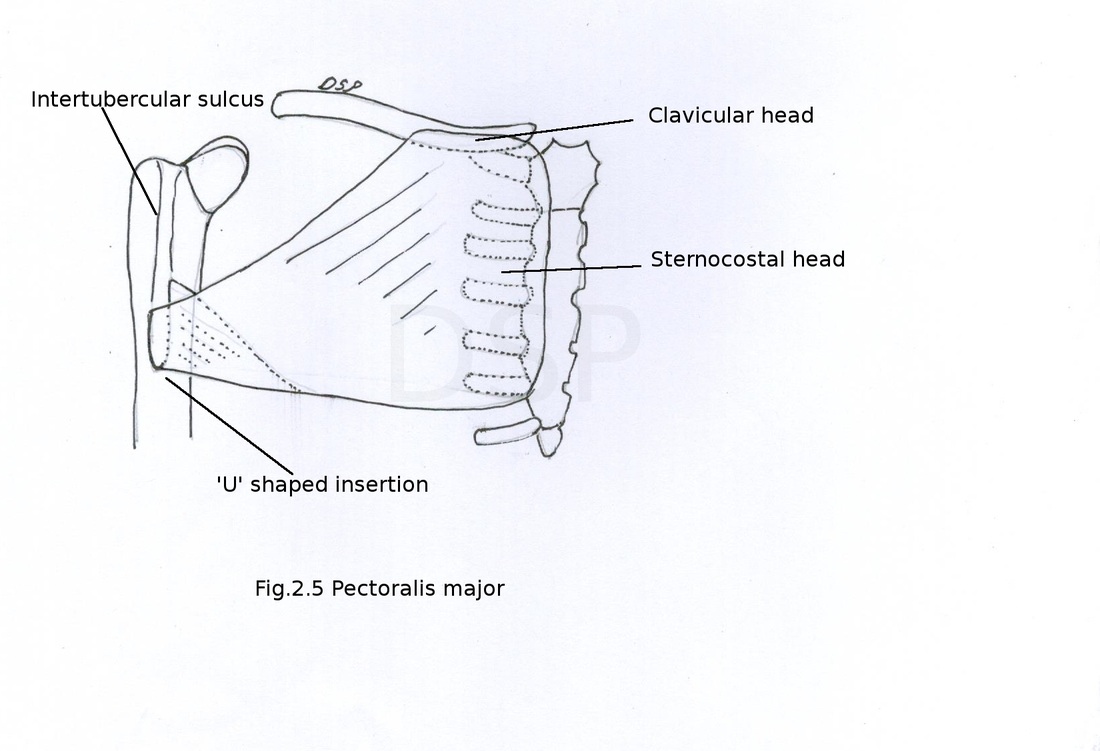
Pectoralis major
Origin: medial ½ of clavicle on anterior surface (clavicular head), anterior part of sternum lateral part up to 6th costal cartilage, 2nd to 6th costal cartilage external oblique aponeurosis (sternocostal head)
Insertion: it forms a bilaminar tendon and shows insertion on lateral lip of inter-tubercular sulcus in 'U' shape manner by two lamina.
Nerve supply: Medial and Lateral pectoral nerves
Action: whole muscle produces Adduction, medial rotation of shoulder joint. Clavicular part shows flexion at shoulder joint. Sternocostal part produce help in climbing by extension of flexed arm. It also help in forced inspiration.
Pectoralis major, Pectoralis minor, Subclavius and part of Serratus anterior.
Pectoralis major
Origin: medial ½ of clavicle on anterior surface (clavicular head), anterior part of sternum lateral part up to 6th costal cartilage, 2nd to 6th costal cartilage external oblique aponeurosis (sternocostal head)
Insertion: it forms a bilaminar tendon and shows insertion on lateral lip of inter-tubercular sulcus in 'U' shape manner by two lamina.
Nerve supply: Medial and Lateral pectoral nerves
Action: whole muscle produces Adduction, medial rotation of shoulder joint. Clavicular part shows flexion at shoulder joint. Sternocostal part produce help in climbing by extension of flexed arm. It also help in forced inspiration.
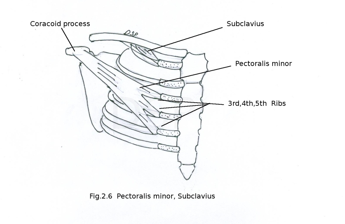
Pectoralis minor
Origin: 3,4, 5th rib near its anterior end and fascia covering external intrcostal muscles.
Insertion: It goes laterally and outwards after forming a tendon shows insertion on medial border and upper surface of coracoid process.
Nerve supply: Medial and Lateral pectoral nerves
Action: Depression of shoulder, protraction of scapula and help in forced inspiration.
Subclavius
Origin: from joining part of 1st rib and its cartilage.
Insertion: goes up and insert on inferior surface of middle 1/3 rd part of clavicle.
Nerve supply: nerve to subclavius branch of upper trunk of brachial plexus
Action: it provide stability to clavicle during movements of upper limb.
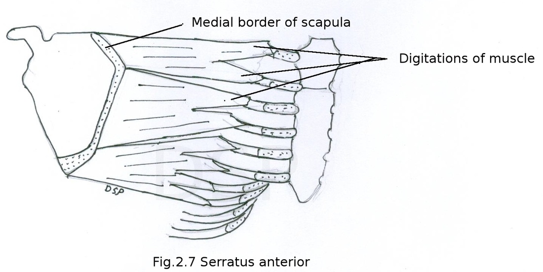
Serratus anterior
Origin: It shows origin from upper 8 ribs by 8 digitations. From fascia covering respective intercostal muscles.
Insertion: Costal surface of scapula 1st digitation – superior angle to root of spine of scapula, 2nd and 3rd digitation - medial border, 4th to 8th digitation- inferior angle of scapula.
Nerve supply: long thoracic nerve from roots of brachial plexus (C5, 6, 7)
Action: projection of scapula for pushing and punching movements, rotation of scapula forward by lower 5 digitations.
