HAND
It shows two surfaces palmar surface and dorsal surface .
Palm
Skin of palm : It is thick firmly fixed to palmar aponeurosis and creased. Numerous sweat glands are present in palmar skin.
Superficial fascia : It bind skin with deep fascia by fibrous band. It contains fat in fibrous compartments.
Deep fascia : It is thickened to form (a) flexor retinaculum, (b) palmar aponeurosis and (c) fibrous flexor sheaths of fingers.
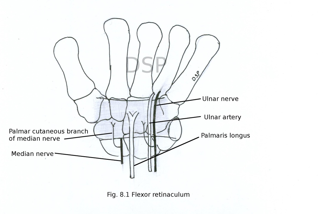
(a) Flexor retinaculum :
It is a strong and fibrous structure which cover anterior or palmar surface of palm and forms carpal tunnel.
Attachments
Medially : hook of hamate, pisiform bone.
Laterally : tubercle of scaphoid and crest of trapezium.
Medially and laterally it shows slip for attachment. Laterally a deep slip which shows attachment on medial lip of a groove on trapezium converting into fibrous tunnel through which tendon of flexor carpi radialis passes. Medially it shows a superficial slip which show attachment on pisiform bone deep to it ulnar vessels and nerve passes.
In distal part it is continuous with palmar aponeurosis and it gives origin to thenar and hypothenar muscles.
Superficial structures
Palmaris longus tendon, palmar cutaneous branch of median nerve, palmar cutaneous branch of ulnar nerve, ulnar nerve and ulnar vessels.
Deep structures
Median nerve, tendon of flexor digitorum superficialis, tendon of flexor digitorum profundus, tendon of flexor pollicis longus, radial bursa and ulnar bursa.
Applied anatomy
Carpal tunnel syndrome : median nerve may get compressed in carpal tunnel. Due to this weakness of thenar muscles and wasting of muscle takes place. Therefore opposition of thumb get affected. Also loss of the cutaneous sensation in palmar surface of lateral 3 and half digits of fingers cause of this maybe arthritis at wrist, carpal joint myxoedema, obesity etc.
It shows two surfaces palmar surface and dorsal surface .
Palm
Skin of palm : It is thick firmly fixed to palmar aponeurosis and creased. Numerous sweat glands are present in palmar skin.
Superficial fascia : It bind skin with deep fascia by fibrous band. It contains fat in fibrous compartments.
Deep fascia : It is thickened to form (a) flexor retinaculum, (b) palmar aponeurosis and (c) fibrous flexor sheaths of fingers.
(a) Flexor retinaculum :
It is a strong and fibrous structure which cover anterior or palmar surface of palm and forms carpal tunnel.
Attachments
Medially : hook of hamate, pisiform bone.
Laterally : tubercle of scaphoid and crest of trapezium.
Medially and laterally it shows slip for attachment. Laterally a deep slip which shows attachment on medial lip of a groove on trapezium converting into fibrous tunnel through which tendon of flexor carpi radialis passes. Medially it shows a superficial slip which show attachment on pisiform bone deep to it ulnar vessels and nerve passes.
In distal part it is continuous with palmar aponeurosis and it gives origin to thenar and hypothenar muscles.
Superficial structures
Palmaris longus tendon, palmar cutaneous branch of median nerve, palmar cutaneous branch of ulnar nerve, ulnar nerve and ulnar vessels.
Deep structures
Median nerve, tendon of flexor digitorum superficialis, tendon of flexor digitorum profundus, tendon of flexor pollicis longus, radial bursa and ulnar bursa.
Applied anatomy
Carpal tunnel syndrome : median nerve may get compressed in carpal tunnel. Due to this weakness of thenar muscles and wasting of muscle takes place. Therefore opposition of thumb get affected. Also loss of the cutaneous sensation in palmar surface of lateral 3 and half digits of fingers cause of this maybe arthritis at wrist, carpal joint myxoedema, obesity etc.
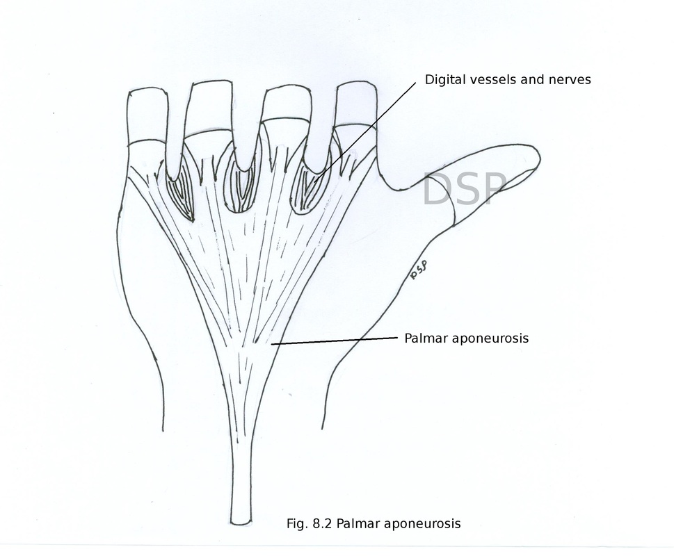
(b) Palmar aponeurosis
It is modification of deep fascia of palm. It shows 3 parts central, medial and lateral. Central part is more thick and prominent medial and lateral parts are thin. Central part is triangular in shape showing apex and base. Apex present slip proximally and join with distal border of flexor retinaculum. Base lies distally and divides into 4 slips. These slips divides into superficial part and deep part which continue with fibrous flexor sheaths. Superficial and deep part shows attachment with superficial transverse ligament of palm and deep transverse ligament of palm, palmar ligaments of metacarpophalangeal joint, base of proximal phalanges respectively. Digital vessels, nerves and tendon of lumbrical muscle passes through interval between slips.
Medial and lateral parts are thin. Medial part shows attachment on shaft of 5th metacarpal bone and lateral part show the attachment on 1st metacarpal bone.
Functions
It improves grip of skin of palm.
It helps in protection of vessels and nerves of palm.
Applied anatomy
Inflammation of palmar aponeurosis : In this condition proximal and middle phalanges are flexed and cannot be extended. Terminal phalanges remains unaffected in this condition ring finger is commonly affected. Name of this condition is dupuytren's contractures.
(c) Fibrous flexor sheaths
Fibrous flexor sheaths of digits are made up for holding tendons of flexor are muscles of digits. It develops from deep fascia of fingers. It shows attachment on sides of phalanges. It extends from head of metacarpal bone to base of distal phalanx. It is thick over phalanges and thin over joints. So it provide a tunnel for flexor tendons of digits.
Fibrous flexor sheaths of digits are made up for holding tendons of flexor are muscles of digits. It develops from deep fascia of fingers. It shows attachment on sides of phalanges. It extends from head of metacarpal bone to base of distal phalanx. It is thick over phalanges and thin over joints. So it provide a tunnel for flexor tendons of digits.
Intrinsic muscles of the hand
Total 20 muscles are present in hand.
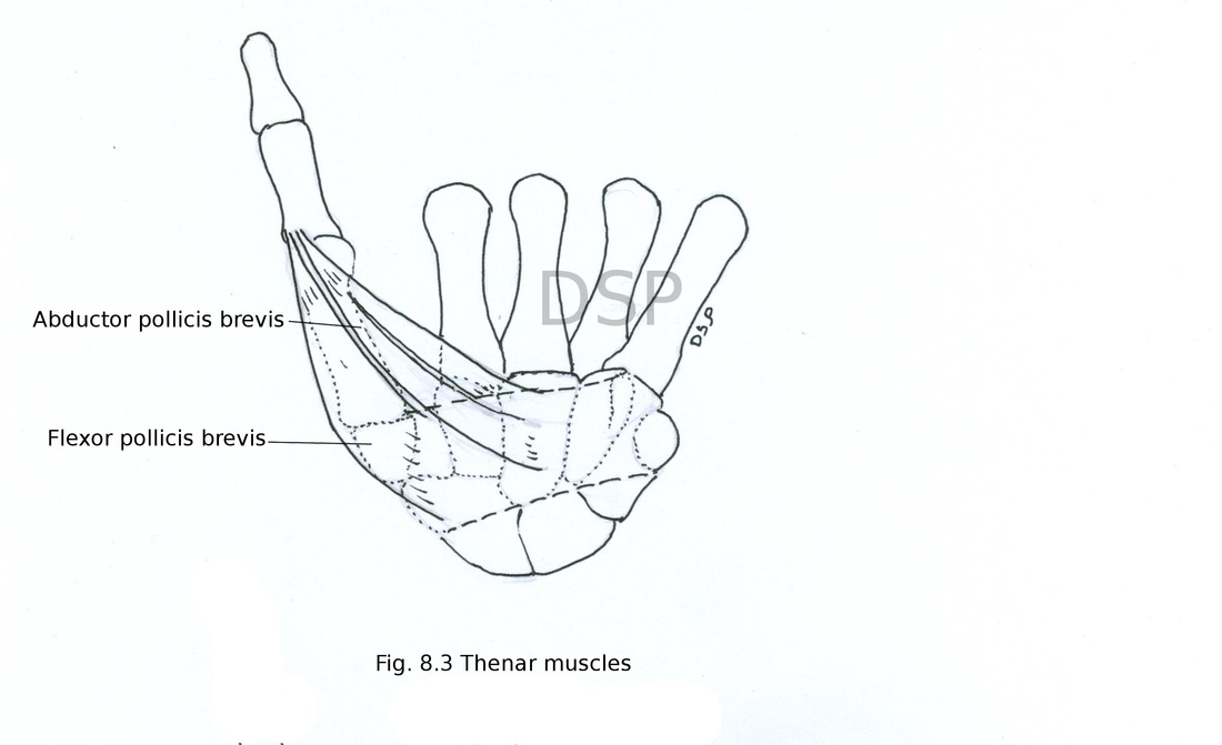
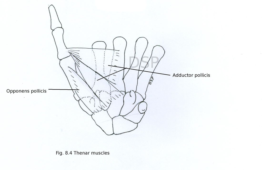
1. Thenar muscles
1. Abductor pollicis brevis
2. Flexor pollicis brevis
3. Opponens pollicis
4. Adductor pollicis
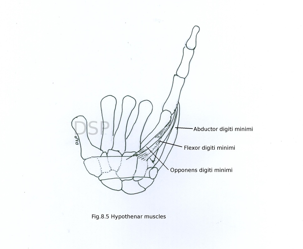
2. Hypothenar muscles
1. Palmaris brevis
2. Abductor digiti minimi
3. Flexor digital minimi
4. Opponens digiti minimi
3. Four lumbricals
4. Four palmar interossei
5. Four dorsal interossei
1. Thenar muscles
1. Abductor pollicis brevis
Origin : It shows origin from flexor retinaculum and tubercles of scaphoid and trapezium.
Insertion: It shows insertion on lateral side of proximal phalanx of thumb and dorsal digital expansion.
Nerve supply from median nerve.
Action : abduction of thumb (rotate thumb medially) at carpo metacarpal and metacarpophalangeal joints.
2. Flexor pollicis brevis
Origin : It shows two heads superficial and deep. Superficial head shows origin from flexor retinaculum and tubercle of trapezium. Deep head shows origin from trapezium and capitate bones.
Insertion : Deep head joins with superficial head and shows insertion on lateral side or radial side of proximal phalanx.
Nerve supply : Superficial head is supplied by recurrent branch of median nerve and deep head from deep branch of a ulnar nerve.
Action : flexion of proximal phalanx thumb.
3. Opponens pollicis
Origin : It shows origin from flexor retinaculum and tubercle of a trapezium.
Insertion : It shows insertion on palmar surface of first metacarpal bone.
Nerve supply : median nerve.
Action : It helps in flexion and medial rotation of first metacarpal bone. Thus it helps in opposition of thumb.
4. Adductor pollicis
Origin : It shows 2 heads of origin oblique and transverse head. Origin of oblique head is from base of second and third metacarpal bone also from capitate and trapezoid. Transverse head shows origin from a verticle line on palmar surface of third metacarpal bone.
Insertion : Both head goes laterally and shows insertion on ulnar or medial side of base of proximal phalanx of thumb.
Nerve supply from the deep branch of ulnar nerve.
Action : It helps in adduction of thumb.
Total 20 muscles are present in hand.
1. Thenar muscles
1. Abductor pollicis brevis
2. Flexor pollicis brevis
3. Opponens pollicis
4. Adductor pollicis
2. Hypothenar muscles
1. Palmaris brevis
2. Abductor digiti minimi
3. Flexor digital minimi
4. Opponens digiti minimi
3. Four lumbricals
4. Four palmar interossei
5. Four dorsal interossei
1. Thenar muscles
1. Abductor pollicis brevis
Origin : It shows origin from flexor retinaculum and tubercles of scaphoid and trapezium.
Insertion: It shows insertion on lateral side of proximal phalanx of thumb and dorsal digital expansion.
Nerve supply from median nerve.
Action : abduction of thumb (rotate thumb medially) at carpo metacarpal and metacarpophalangeal joints.
2. Flexor pollicis brevis
Origin : It shows two heads superficial and deep. Superficial head shows origin from flexor retinaculum and tubercle of trapezium. Deep head shows origin from trapezium and capitate bones.
Insertion : Deep head joins with superficial head and shows insertion on lateral side or radial side of proximal phalanx.
Nerve supply : Superficial head is supplied by recurrent branch of median nerve and deep head from deep branch of a ulnar nerve.
Action : flexion of proximal phalanx thumb.
3. Opponens pollicis
Origin : It shows origin from flexor retinaculum and tubercle of a trapezium.
Insertion : It shows insertion on palmar surface of first metacarpal bone.
Nerve supply : median nerve.
Action : It helps in flexion and medial rotation of first metacarpal bone. Thus it helps in opposition of thumb.
4. Adductor pollicis
Origin : It shows 2 heads of origin oblique and transverse head. Origin of oblique head is from base of second and third metacarpal bone also from capitate and trapezoid. Transverse head shows origin from a verticle line on palmar surface of third metacarpal bone.
Insertion : Both head goes laterally and shows insertion on ulnar or medial side of base of proximal phalanx of thumb.
Nerve supply from the deep branch of ulnar nerve.
Action : It helps in adduction of thumb.
Hypothenar muscles
These are small muscles present in the hypothenar eminence on medial side of hand.
1. Palmaris brevis
Origin : It shows origin from flexor retinaculum and palmar aponeurosis.
Insertion : Muscle shows insertion on skin of medial border of hand.
Nerve supply from superficial branch of ulnar nerve.
Action : It helps in gripping by hand.
2. Abductor digiti minimi
Origin : It shows origin from palmar aspect of pisiform bone, continuation of tendon of flexor carpi ulnaris and pisohamate ligament.
Insertion : It shows insertion on ulnar side of base of proximal phalanx of little finger.
Nerve supply from the deep branch of ulnar nerve.
Action : Abduction of little finger.
3. Flexor digiti minimi
Origin : It shows origin from flexor retinaculum and hook of hamate.
Insertion : It shows insertion on proximal phalanx of little finger near its base and on ulnar side.
Nerve supply ulnar nerve provides its deep branch.
Action : flexion at metacarpophalangeal joint of little finger.
4. Opponens digiti minimi
Origin : It shows origin from flexor retinaculum and hook of hamate.
Insertion : It shows insertion on palmar surface of ulnar side of fifth metacarpal bone.
Nerve supply ulnar nerve by provide its deep branch.
Action : Flexion of fifth metacarpal bone.
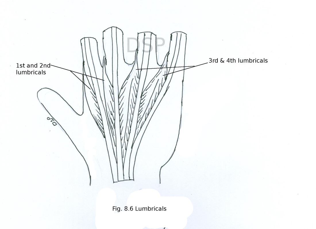
Lumbricals
These are four small muscles of hand numbered from lateral to medial side these muscles takes origin from tendon of flexor digitorum profundus and joins with dorsal digital expansion.
First and second lumbricals are unipennate muscles while third and fourth lumbricals are bipennate muscles.
Origin :
First lumbrical : It shows origin from radial side of profundus tendon for index finger.
Second lumbrical : It shows origin from radial side of profundus tendon for middle finger.
Third lumbrical : It shows origin from both sides of profundus tendons for middle and ring fingers.
Fourth lumbrical : It shows origin from both sides of profundus tendons for ring and little fingers.
Insertion :
First second third and fourth lumbricals goes backward on radial side of respective fingers shows insertion on dorsal digital expansion of that finger.
Nerve supply : First and second lumbricals are supplied by median nerve. Third and fourth lumbricals receive nerve supply from ulnar nerve by it's deep branch.
Action : Lumbricals help in flexion of digit at metacarpophalangeal joint and extension at interphalangeal joint.
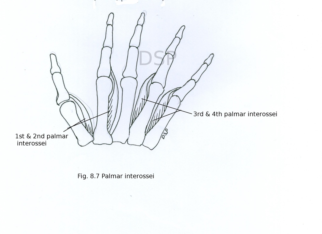
Palmar interossei
Palmar interossei are four small muscles present on palmar aspect of hand. These are numbered from lateral to medial side.
Origin :
First palmar interosseous muscle shows origin from ulnar side of base of first metacarpal bone.
Second palmar interosseous muscle shows origin from ulnar side of shaft of palmar aspect of second metacarpal bone.
Third Palmar interosseous muscle shows origin from radial side of palmar aspect of shaft of fourth metacarpal bone.
Fourth Palmar interosseous muscle shows origin from radial side of palmar aspect of shaft of fifth metacarpal bone.
Insertion :
All interosseous muscles shows insertion on dorsal digital expansion of respective fingers also shows insertion on base of proximal phalanx of respective fingers on the same side of their origin.
First palmar interosseous muscle shows insertion on ulnar side of proximal phalanx of thumb.
Second palmar interosseous muscle shows insertion on ulnar side of proximal phalanx of index finger .
Third palmar interosseous muscle shows insertion on radial side of proximal phalanx of 4th finger.
Fourth palmar interosseous muscle shows insertion on radial side of proximal phalanx of fifth finger.
Actions : palmar interosseous help in adduction of fingers. It also helps in flexion at metacarpophalangeal joint and extension at interphalangeal joints.
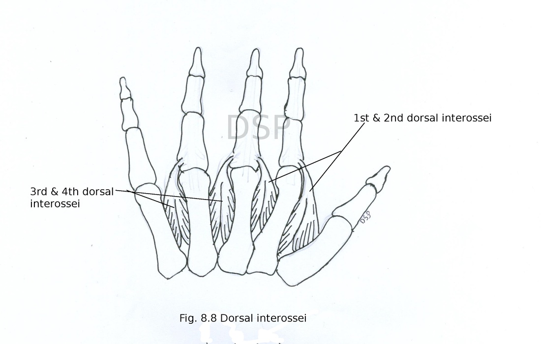
Dorsal interossei
Origin :
Dorsal interossei are bipennate muscles numbered from lateral to medial side. These are 4 in number.
First dorsal interosseous shows origin from shafts of first and second metacarpal bones on palmar surface.
Second dorsal interosseous muscle shows origin from shafts of second and third metacarpal bone on palmar surface.
Third dorsal interosseous muscle shows origin from shafts of third and fourth metacarpal bones on palmar surface.
Fourth dorsal interosseous muscle shows origin from shafts of 4th and 5th metacarpal bones on palmar surface.
Insertion :
First dorsal interosseous muscle shows insertion on radial side of base of proximal phalanx of index finger and also join with dorsal digital expansion of same finger.
Second dorsal interosseous muscle shows insertion on radial side of base of proximal phalanx of middle finger also join with dorsal digital expansion of the same finger.
Third dorsal interosseous muscle shows insertion on a ulnar side of base of proximal phalanx of middle finger also joins with dorsal digital expansion of the same finger.
Fourth dorsal interosseous muscle shows insertion on ulnar side of base of proximal phalanx of ring finger also shows insertion on dorsal digital expansion of the same finger.
Nerve supply dorsal interossei receives nerve supply from ulnar nerve by its deep branch.
Action : dorsal interossei helps in abduction of 2nd, 3rd and 4th fingers. It also helps in flexion at metacarpophalangeal joint and extension and intephalangeal joints.
Arteries in the hand
Radial and ulnar artery in hand anastomose with each other and form two palmar arches.
(1) Superficial palmar arch and (2) Deep palmar arch.
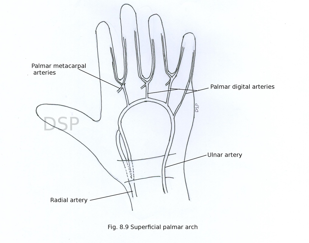
(1) Superficial palmar arch
Superficial palmar arch is formed by ulnar artery present in the palm. Enters in palmar surface of hand superficial to flexor retinaculum and it is completed on lateral side by any one of the following arteries superficial palmar branch of the radial artery, arteria princeps pollicis, arteria radialis indicis. It is convex distally. This arch lies deep to palmar aponeurosis and anterior to long flexor tendons of hand, lumbricals muscles and digital branches of median nerve.
branches
4 palmar digital arteries arise from convex side of superficial palmar arch. First palmar digital artery runs along ulnar side of fifth finger while remaining three arteries run as common palmar digital arteries which joins with palmar metacarpal arteries of deep palmar arch. Finally it supply sides of respective fingers by dividing into branches.
Radial and ulnar artery in hand anastomose with each other and form two palmar arches.
(1) Superficial palmar arch and (2) Deep palmar arch.
(1) Superficial palmar arch
Superficial palmar arch is formed by ulnar artery present in the palm. Enters in palmar surface of hand superficial to flexor retinaculum and it is completed on lateral side by any one of the following arteries superficial palmar branch of the radial artery, arteria princeps pollicis, arteria radialis indicis. It is convex distally. This arch lies deep to palmar aponeurosis and anterior to long flexor tendons of hand, lumbricals muscles and digital branches of median nerve.
branches
4 palmar digital arteries arise from convex side of superficial palmar arch. First palmar digital artery runs along ulnar side of fifth finger while remaining three arteries run as common palmar digital arteries which joins with palmar metacarpal arteries of deep palmar arch. Finally it supply sides of respective fingers by dividing into branches.
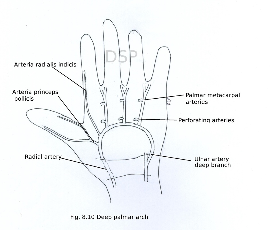
(2) Deep palmar arch
Deep palmar arch is formed by anastomosis of terminal branch of radial artery and deep branch of ulnar artery. It lies deep to long flexor tendons of hand. Radial artery enter in palm after passing between oblique and transverse heads of adductor pollicis to continue as deep palmar arch. During its course radial artery gives branches arteria princep pollicis and arteria radialis indices. Arteria princep pollicis supply 2 sides of thumb while arteria radialis indicis supply radial side of index finger. Deep palmar arch lies deep to oblique head of adductor pollicis, long flexor tendons and lumbrical muscles.
Branches
3 palmar metacarpal arteries passes distally in second, third and fourth spaces. Finally anastomoses with common palmar metacarpal branches of superficial palmar arch.
3 perforating arteries after passing between dorsal interosseous muscle joins with dorsal metacarpal arteries.
Recurrent branches are in relation with carpal bones and anastomose with palmar carpal arch.
Nerves of the hand
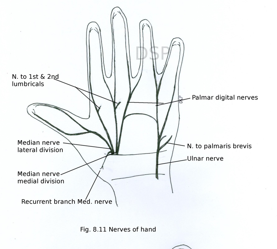
Median nerve
Median nerve enters in the palm of hand deep to flexor retinaculum. Then it divides into two branches lateral and medial. It gives out a muscular recurrent branch to supply from lateral side to supply 3 thenar muscles abductor pollicis brevis, flexor pollicis brevis and opponens pollicis.
Lateral branch: It gives 3 palmar digital nerves for lateral one and half fingers to supply skin of both sides of thumb and radial side of index finger. Branch to index finger give out a small muscular branch to supply first lumbrical muscle.
Medial branch : It is divided into 2 palmar digital nerves to supply ulnar side of index finger and radial side of middle finger, ulnar side of the middle finger and radial side of a ring finger. Digital branch which supply adjacent sitds of index and middle finger also gives a muscular branch to supply 2nd lumbrical muscle.
Therefore median nerve supply lateral three and half fingers skin, three thenar muscles, first and second lumbrical muscles.
Ulnar nerve
Ulnar nerve appears in palmar surface of hand by passing superficial to flexor retinaculum and on lateral side of pisiform bone. Here it divides into 2 branches superficial terminal branch and deep terminal branch.
Superficial terminal branch gives a small branch to palmaris brevis muscle and then it gives out two palmar cutaneous branches to supply ulnar side of little finger and a lateral common palmar digital branch to radial side of the little and ulnar side of ring finger.
Deep terminal branch passes deep in between origin of abductor digiti minimi and flexor digiti minimi passes laterally after piercing opponens digiti minimi muscle in relation with below the hook of hamate alongwith concavity of deep palmar arch and deep 2 long flexor tendons of hand. During its course supply 3 hypothenar muscles, 3rd and 4th lumbrical muscles, 4 dorsal and 4 palmar interossei also a branch to adductor pollicis and deep head of flexor pollicis brevis.
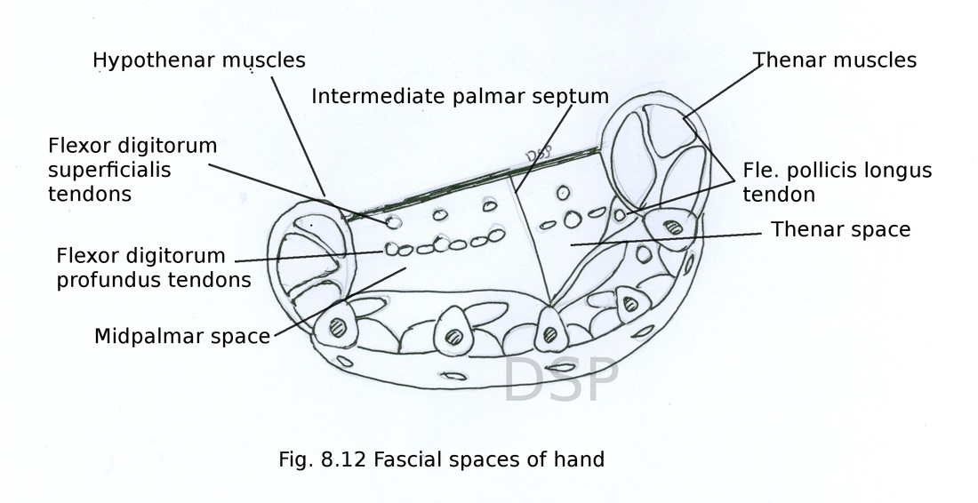
Fascial spaces of hand
Because of arrangement of fascia spaces are created in hand. These spaces are surgically important because infection and collection of pus may takes place.
Mid palmar space
It is triangular shaped space in hand.
Boundaries
Anteriorly : long flexor tendons of 3rd, 4th and 5th fingers, third and fourth lumbrical muscles and palmar aponeurosis.
Posteriorly : Third and fourth metacarpal spaces with interossei and metacarpal bones of that space.
Laterally : Intermediate fibrous septum.
Medially : Medial palmar septum covering hypothenar muscles.
Proximally it extends from distal margin of flexor retinaculum to distally up to the space of fingers between third and fourth fingers.
Thenar space
It is a triangular shaped space present in hand.
Anteriorly : Thenar muscles, long flexor tendon of index finger, first and second lumbrical muscles and palmar aponeurosis.
Posteriorly : Adductor pollicis muscle, 1st dorsal interosseous muscle.
Laterally : Lateral palmar septum with tendon of flexor pollicis longus.
Medially : Intermediate fibrous septum.
It extends proximally from distal border of flexor retinaculum to distally space between fingers on both side of index finger.
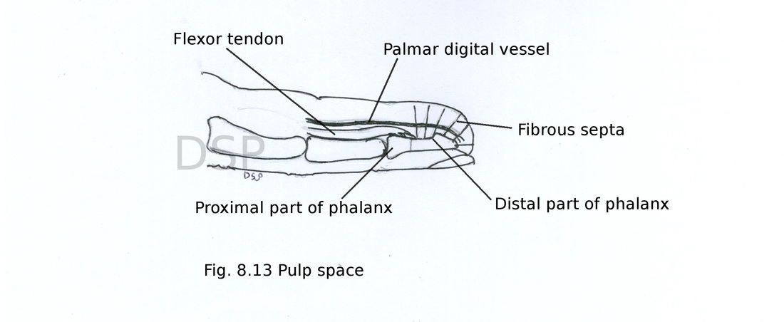
Pulp space of finger
These are spaces present in distal phalanges of hand. Distal phalanges of hand is divided into compartments containing fat and blood vessels. The compartments are formed by fibrous septa which connect palmar skin and periosteum of distal phalanx. These spaces are present distal to attachment of long flexor tendon on distal phalanx.
Applied : Infection of compartments may takes place with formation of pus. For drainage lateral incision is must. Name of this condition is whitlow. If not treated early it may cause avascular necrosis of distal four fifth part of terminal phalanx but proximal one fifth part remains unaffected cause is artery not present in fibrous septa in this region.