Lateral side of Leg ( peroneal compartment)
Lateral compartment of leg is bounded by
Anteriorly : Anterior intermuscular septum
Posteriorly : Posterior intermuscular septum
Medially : Lateral surface of fibula
Laterally : Deep fascia of leg
Contents
Muscles : Peroneus longus and peroneus brevis
Vessels : Peroneal branch of posterior tibial artery
Nerve : Superficial peroneal nerve
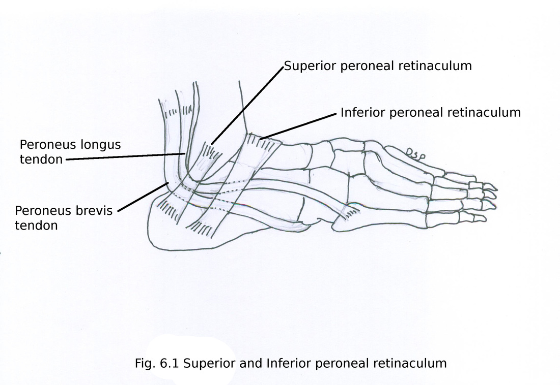
Near lower part of leg on lateral aspect of ankle joint deep fascia get modified into superior and inferior peroneal retinaculum.
Superior peroneal retinaculum
It is a band of deep fascia present on lateral aspect of lower part of leg in relation with ankle joint.
Attachments : Anteriorly it shows attachment on posterior margin of lateral malleolus of fibula and posteriorly it shows attachment on lateral surface of calcaneum also on deep transverse fascia of leg. It enclose tendon of peroneus longus and peroneus brevis muscles.
Inferior peroneal retinaculum
It is in continuity with inferior extensor retinaculum.
Attachments : Above shows attachment on anterior part of superior surface of calcaneus and peroneal trochlea. Below it shows attachment on lateral surface of calcaneus.
Attachment on peroneal trochlea forms two compartments. Through upper part passes peroneus brevis and through lower part passes peroneus longus.
Lateral compartment of leg is bounded by
Anteriorly : Anterior intermuscular septum
Posteriorly : Posterior intermuscular septum
Medially : Lateral surface of fibula
Laterally : Deep fascia of leg
Contents
Muscles : Peroneus longus and peroneus brevis
Vessels : Peroneal branch of posterior tibial artery
Nerve : Superficial peroneal nerve
Near lower part of leg on lateral aspect of ankle joint deep fascia get modified into superior and inferior peroneal retinaculum.
Superior peroneal retinaculum
It is a band of deep fascia present on lateral aspect of lower part of leg in relation with ankle joint.
Attachments : Anteriorly it shows attachment on posterior margin of lateral malleolus of fibula and posteriorly it shows attachment on lateral surface of calcaneum also on deep transverse fascia of leg. It enclose tendon of peroneus longus and peroneus brevis muscles.
Inferior peroneal retinaculum
It is in continuity with inferior extensor retinaculum.
Attachments : Above shows attachment on anterior part of superior surface of calcaneus and peroneal trochlea. Below it shows attachment on lateral surface of calcaneus.
Attachment on peroneal trochlea forms two compartments. Through upper part passes peroneus brevis and through lower part passes peroneus longus.
Muscles :
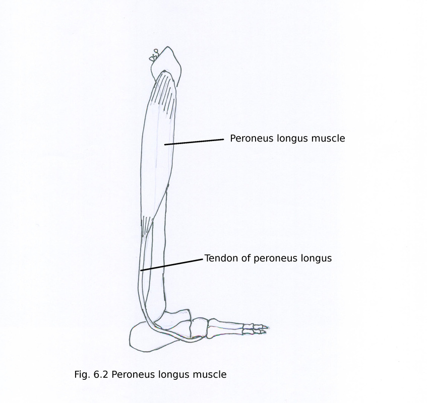
Peroneus longus
It is a bipennate in upper part and unipennate in lower part.
Origin : It shows origin from head, upper two third part of lateral surface of fibula , part of lateral condyle tibia, deep surface of deep fascia, anterior and posterior intermuscular septum.
Insertion : Below in lower part of leg this muscle forms a tendon which lies superficial to tendon of peroneus brevis. Tendon passes through superior peroneal retinaculum along with tendon of peroneus brevis and in inferior peroneal retinaculum it lies in lower compartment. It goes medially and forward in relation to cuboid. After passing through osseo-fibrous tunnel on plantar aspect of cuboid bone shows insertion on infero lateral surface of first metatarsal bone and adjacent surface of medial cuneiform bone, a small slip on second metatarsal bone.
Nerve supply : It receives nerve supply from superficial peroneal nerve L5, S1
Action : Evertor of foot, maintain lateral longitudinal arch from above, maintain transverse arch of foot. It also plantar flex ankle joint.
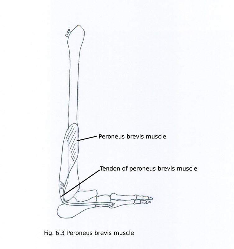
Peroneus brevis
It is a bipennate muscle.
Origin : It shows origin from lower two third part of lateral surface of shaft of fibula anterior to peroneus longus, anterior and posterior intermuscular septum.
Insertion : Below in lower part of leg this muscle forms a tendon. Tendon passes deep to medial malleolus in superior peroneal retinaculum. Then it goes downward through upper part of inferior peroneal retinaculum. It shows insertion on tubercle on lateral aspect of base of fifth metatarsal bone.
Nerve supply : It receives nerve supply from superficial peroneal nerve L5, S1.
Action : Evertor of foot, maintain lateral longitudinal arch from above.
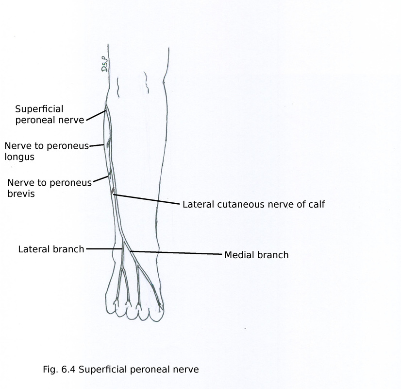
Nerve :
Superficial peroneal nerve (musculo-cutaneous nerve) L4, L5, S1
It is a branch of common peroneal nerve. It arise near neck of fibula deep to peroneus longus. Then it goes downward between peroneus longus and brevis. Below at the junction of upper two third and lower one third of leg it pierces deep fascia of leg. Here it gives cutaneous branch to lower part of leg and divides into two branches medial and lateral. Medial and Lateral branch supply skin of dorsum of foot.
Branches :
1) Muscular branches : It supply muscles peroneus longus and brevis.
2) Cutaneous branches : Medial branch gives two dorsal digital nerves one for medial aspect of great toe and second branch for adjacent side of second and third toe. Lateral branch gives two dorsal digital nerve to supply adjacent sides of third to fifth toes.