Anterior compartment of Thigh and Adductor region
Skin
It is thick and fold of groin present on its upper part.
Superficial fascia
It consists of two layers in upper part superficial fatty and deep membranous layer in relation with anterior abdominal wall. Superficial layer continue with Campers fascia of abdominal wall. Deep layer forms cribriform fascia.
It contain fat, cutaneous nerves, great saphenous vein, cutaneous arteries, lymph nodes and lymph vessels.
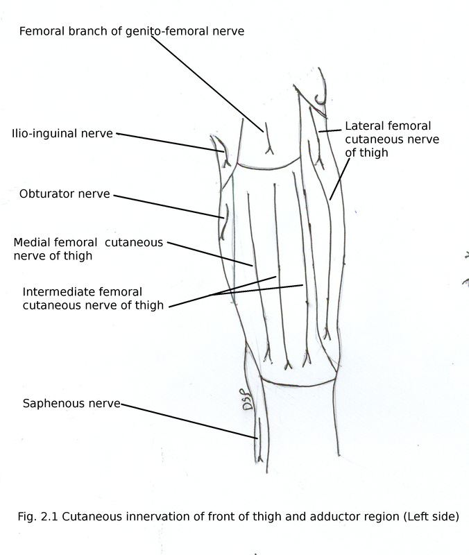
Cutaneous nerves
Skin of front of thigh supplied by cutaneous nerves derived from lumbar plexus.
1) Lateral femoral cutaneous nerve L2 L3
It is a branch of lumbar plexus. It divides into two branches anterior and posterior. Supply anterior and lateral part of upper part of thigh.
2) Intermediate femoral cutaneous nerve L2 L3
It is a branch of anterior division of femoral nerve. Pierces fascia lata near middle part of sartorius muscle. After dividing into branches supply skin of front of thigh middle part upto knee.
3) Medial femoral cutaneous nerve L2 L3
It is a branch of anterior division of femoral nerve. Pierces fascia lata near lower part of sartorius muscle. It divides into two branches anterior and posterior. Supply skin of medial part of thigh.
4) Branch of Obturator nerve L2 L3 L4
Anterior division of obturator nerve supply medial and lower part of thigh.
5) Ilioinguinal nerve L1
It is a branch of lumbar plexus. It comes out through superficial inguinal ring. Supply skin of medial part of upper aspect of thigh, root of penis, upper part of scrotum in male and mons pubis and labium majus in female.
6) Femoral branch of genitofemoral nerve L1 L2
It is a branch of lumbar plexus. After piercing femoral sheath supply skin of femoral triangle.
Skin
It is thick and fold of groin present on its upper part.
Superficial fascia
It consists of two layers in upper part superficial fatty and deep membranous layer in relation with anterior abdominal wall. Superficial layer continue with Campers fascia of abdominal wall. Deep layer forms cribriform fascia.
It contain fat, cutaneous nerves, great saphenous vein, cutaneous arteries, lymph nodes and lymph vessels.
Cutaneous nerves
Skin of front of thigh supplied by cutaneous nerves derived from lumbar plexus.
1) Lateral femoral cutaneous nerve L2 L3
It is a branch of lumbar plexus. It divides into two branches anterior and posterior. Supply anterior and lateral part of upper part of thigh.
2) Intermediate femoral cutaneous nerve L2 L3
It is a branch of anterior division of femoral nerve. Pierces fascia lata near middle part of sartorius muscle. After dividing into branches supply skin of front of thigh middle part upto knee.
3) Medial femoral cutaneous nerve L2 L3
It is a branch of anterior division of femoral nerve. Pierces fascia lata near lower part of sartorius muscle. It divides into two branches anterior and posterior. Supply skin of medial part of thigh.
4) Branch of Obturator nerve L2 L3 L4
Anterior division of obturator nerve supply medial and lower part of thigh.
5) Ilioinguinal nerve L1
It is a branch of lumbar plexus. It comes out through superficial inguinal ring. Supply skin of medial part of upper aspect of thigh, root of penis, upper part of scrotum in male and mons pubis and labium majus in female.
6) Femoral branch of genitofemoral nerve L1 L2
It is a branch of lumbar plexus. After piercing femoral sheath supply skin of femoral triangle.
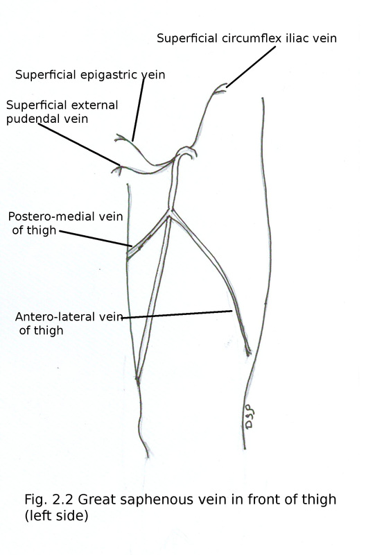
Veins
Great saphenous vein
After its long course from dorsum of foot, posterior aspect of knee goes upwards on medial side of thigh and reach saphenous opening passes through it and drains into femoral vein by piercing cribriform fascia.
Tributaries of great saphenous vein are in thigh :
1) Antero-lateral vein of thigh from anterior part of thigh.
2) Postero-medial vein of thigh from posterior and medial part of thigh.
Three superficial veins alongwith three superficial branches of femoral artery.
3) Superficial epigastric
4) Superficial circumflex iliac
5) Superficial external pudendal vein
Tributaries before opening in to femoral vein
6) Deep external pudendal vein
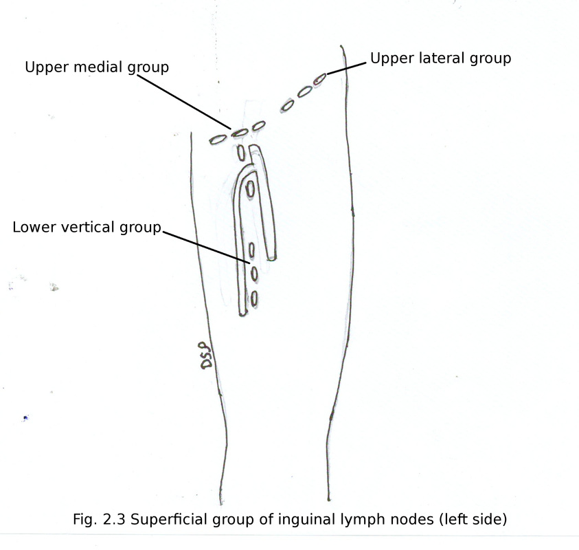
Lymph nodes
Inguinal group of lymph nodes are present in this region in two groups 1) superficial and 2) deep.
1) Superficial group arranged into upper horizontal and lower vertical group. Upper group is divided into lateral and medial group. Upper group lies below inguinal ligament. Upper lateral group drains from antero-lateral and lower part of anterior abdominal wall, gluteal region. Upper medial group drains from antero-medial and lower part of anterior abdominal wall, penis in male, vulva and vagina in female, from angle of uterus near attachment of uterine tube, perineum, lower part of anal canal. Lower vertical group lies in relation with lateral part of great saphenous vein near termination drains lower limb below it. All these nodes drains into external iliac nodes.
2) Deep inguinal group of lymph nodes lies in relation with femoral vein draining lymph from vessels which runs along with branches of femoral vessels, glans penis in male, glans clitoris in female and from superficial inguinal group of lymph nodes.
Applied anatomy of inguinal lymph nodes:
Swelling or enlargement of inguinal group of lymph nodes occurs due to malignancy or diseases in lower limb, anterior abdominal wall below umbilicus, gluteal region, penis or clitoris and lower part of anal canal.
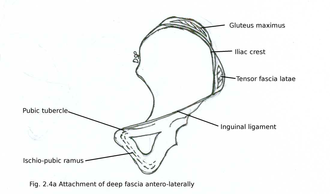
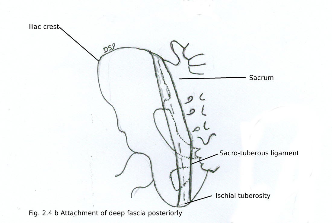
Deep fascia of thigh
Fascia lata is deep fascia of thigh. Fascia lata shows attachment on back of sacrum and coccyx posteriorly. Antero-laterally attached on outer margin of iliac crest then anteriorly on inguinal ligament, superior ramus of pubis continue downward on inferior ramus of pubis, ramus of ischium and tuberosity of ischium then continue backwards on lower margin of sacrotuberous ligament.
It splits to inclose gluteus maximus and tensor fascia latae superficial and deep to the muscles respectively.
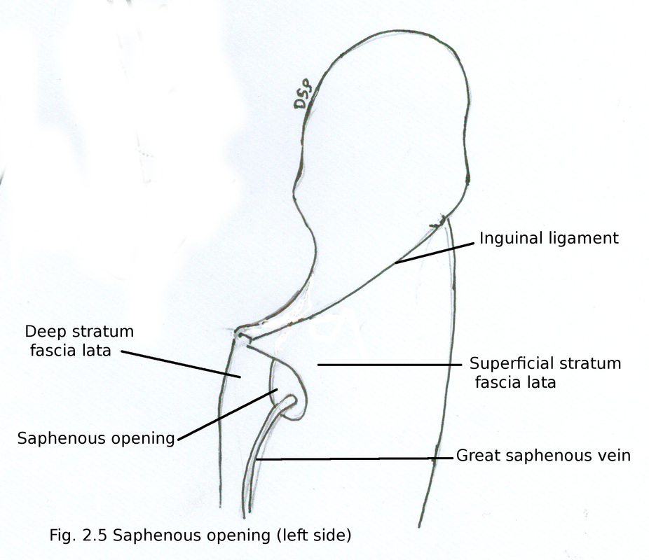
Saphenous opening
It is an opening in anterior aspect of thigh in deep fascia. It is inferolateral to pubic tubercle about 3cm. Size of it is about 3-4 cm. Deep fascia shows two strata in relation to opening superficial and deep. Superficial stratum shows attachment on inguinal ligament, crest and anterior superior iliac spine of ilium, pecten pubis. It shows reflexion inferolaterally forming superior, lateral and inferior margins. Deep stratum forms medial margin of opening goes upwards and shows attachment on pecten pubis. Saphenous opening covered by cribriform fascia.
Structures which passes through saphenous opening :
Great saphenous vein, superficial epigastric artery, superficial external pudendal artery, lymph vessels connecting superficial and deep inguinal group of lymph nodes.
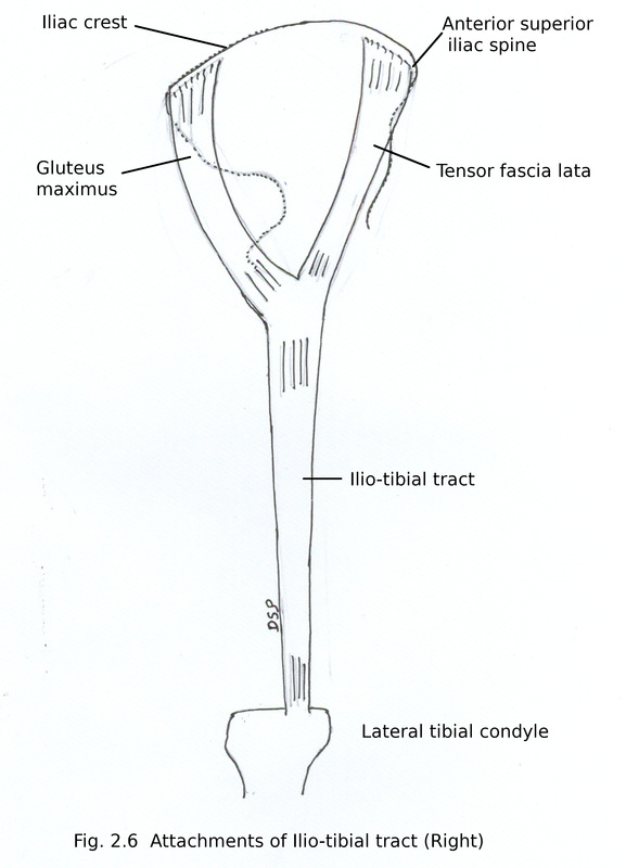
Ilio-tibial tract
It is thick band of fascia lata on lateral surface of thigh. In upper part it splits into two layers for insertion of tensor fascia lata and posteriorly shows insertion of 3/4th part of gluteus maximus. Superficial layer after crosssing gluteus maximus superficially shows attachment on iliac crest and deep layer passes deep to gluteus maximus muscle shows attachment on capsule of hip joint. Below continue as a band and shows attachment on triangular area on antero-lateral aspect of lateral condyle of tibia.
Functions of ilio-tibial tract is to stabilize knee joint in extension and semiflexion. Supports during walking and running.
Intermuscular septum
Fascia lata gives out two intermuscular septum lateral and medial. Showing attachment on linea aspera. Lateral septum present in between vastus lateralis and short head of biceps femoris. Medial septum present in between vastus medialis and pectineus alonwith adductor muscles of thigh.
Other structures of front of thigh
Other structures of front of thigh lies deep to fascia lata. These are femoral triangle, adductor canal, part of adductor region and quadriceps group of muscles.
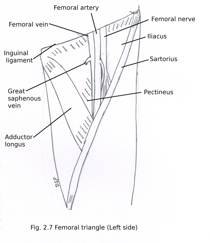
Femoral Triangle
It is triangular depressed area below inguinal ligament in upper part of front of thigh.
Boundaries :
Laterally medial border of sartorius muscle .
Medially medial border of adductor longus.
Base inguinal ligament.
Apex formed by sartorius and adductor longus where it meets below.
Floor formed by iliacus, tendon of psoas major, pectineus and adductor longus from lateral to medial side.
Roof formed by fascia lata.
Contents :
1. Femoral artery and its branches
2. Femoral vein and its tributaries
3. Femoral nerve and its branches
4. Deep inguinal group of lymph nodes
5. Lateral femoral cutaneous nerve
6. Femoral branch of genitofemoral nerve
Fascia lata gives out two intermuscular septum lateral and medial. Showing attachment on linea aspera. Lateral septum present in between vastus lateralis and short head of biceps femoris. Medial septum present in between vastus medialis and pectineus alonwith adductor muscles of thigh.
Other structures of front of thigh
Other structures of front of thigh lies deep to fascia lata. These are femoral triangle, adductor canal, part of adductor region and quadriceps group of muscles.
Femoral Triangle
It is triangular depressed area below inguinal ligament in upper part of front of thigh.
Boundaries :
Laterally medial border of sartorius muscle .
Medially medial border of adductor longus.
Base inguinal ligament.
Apex formed by sartorius and adductor longus where it meets below.
Floor formed by iliacus, tendon of psoas major, pectineus and adductor longus from lateral to medial side.
Roof formed by fascia lata.
Contents :
1. Femoral artery and its branches
2. Femoral vein and its tributaries
3. Femoral nerve and its branches
4. Deep inguinal group of lymph nodes
5. Lateral femoral cutaneous nerve
6. Femoral branch of genitofemoral nerve
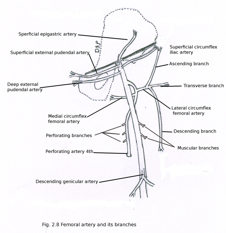
1. Femoral artery
It is a continuation of external iliac artery. It passes deep to inguinal ligament midway between anterior superior iliac spine and pubic symphysis. It passes through femoral triangle from base to apex then enters inside adductor canal then continue as popliteal artery in popliteal fossa after passing through opening in adductor magnus. Proximal part of femoral artery near about 4cm is present inside femoral sheath.
Branches:
A. Superficial epigastric artery :
Arises below inguinal ligament. It goes upwards in superficial fascia of anterior abdominal wall and supply lower part of anterior abdominal wall, superficial inguinal lymph nodes and skin around it.
B. Superficial circumflex iliac artery :
Arises along with superficial epigastric artery. Goes laterally just below inguinal ligament towards anterior superior iliac spine. Forms spinous anastomosis with deep circumflex femoral artery, deep branch of superior gluteal and ascending branch of lateral circumflex femoral arteries.
C. Superficial external pudendal artery :
After passing through cribriform fascia goes medially. It supply skin of scrotum in male or labium majus in female.
D. Deep external pudendal artery :
Goes medially to supply scrotum in male and labium majus in female. It anastomose with posterior scrotal or labial branches of internal pudendal artery.
E. Muscular branches :
Supply muscles adductors, vastus medialis and sartorius.
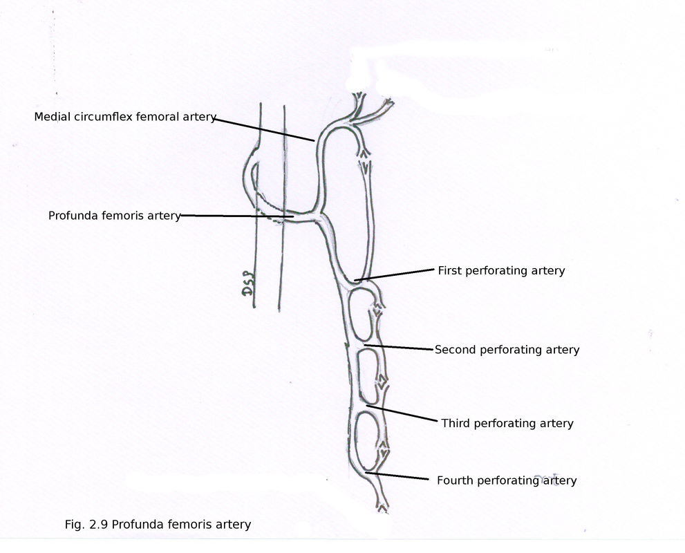
F. Profunda femoris :
It is a big branch of femoral artery. It is present on lateral aspect of femoral artery about 3.5 cm below inguinal ligament. Then it goes posterior to femoral artery. Then it passes between pectineus and adductor longus, adductor longus and brevis respectively. Finally descends between adductor longus and magnus. After piercing adductor magnus continue as 4th perforating artery to anastomose with upper muscular branch of popliteal artery.
Branches:
a) Lateral circumflex femoral artery :
Goes laterally between two branches of femoral nerve. Passes deep to sartorius and divides into three branches ascending , descending and transverse.
Ascending branch goes up towards anterior superior iliac spine. Anastomose with superior gluteal, deep circumflex iliac, superficial circumflex iliac, branch of medial circumflex iliac. (spinous anastomosis).
Descending branch goes downwards along anterior margin of vastus lateralis. Anastomose with superior lateral genicular branch of popliteal artery (anastomosis around knee joint).
Transverse branch goes laterally by piercing vastus lateralis and below greater trochanter anastomose with medial circumflex femoral, inferior gluteal and first perforating artery (cruciate anastomosis).
b) Medial circumflex femoral artery :
It goes medially between psoas major and pectineus, adductor brevis and obturator externus, quadratus femoris and upper margin of adductor magnus respectively. It divides into transverse and descending branch.
Transverse branch forms cruciate anastomosis.
Descending branch goes to trochanteric fossa and anastomose with inferior branch of superior gluteal artery, descending branch of inferior gluteal artery, ascending branch of lateral circumflex femoral artery (trochanteric anastomosis). An acetabular branch supply acetabular fat and head of femur near ligamentum teres.
c) Perforating artery :
These are four but fourth is actually continuation of profunda femoris. It supply adductor and hamstring muscles. First branch passes above adductor brevis, second in front of adductor brevis and third below adductor brevis. Nutrient artery for femur are branches of first and second perforating. It gives out ascending and descending branches to anastomose with branches of each other and upper most ascending branch anastomose for cruciate anastomosis. Lower most descending branch anastomose with muscular branches of popliteal artery.
d) Muscular artery :
It gives out branches to adductors, flexor muscles, and anastomose with medial circumflex femoral and muscular branches from popliteal artery.
G. Descending genicular artery :
It arises in adductor canal and gives out a saphenous branch. Goes downwards in the substance of vastus medialis. Anastomose with medial superior genicular artery. Saphenous branch runs along with saphenous nerve and anastomose with medial inferior genicular artery.
It is a continuation of external iliac artery. It passes deep to inguinal ligament midway between anterior superior iliac spine and pubic symphysis. It passes through femoral triangle from base to apex then enters inside adductor canal then continue as popliteal artery in popliteal fossa after passing through opening in adductor magnus. Proximal part of femoral artery near about 4cm is present inside femoral sheath.
Branches:
A. Superficial epigastric artery :
Arises below inguinal ligament. It goes upwards in superficial fascia of anterior abdominal wall and supply lower part of anterior abdominal wall, superficial inguinal lymph nodes and skin around it.
B. Superficial circumflex iliac artery :
Arises along with superficial epigastric artery. Goes laterally just below inguinal ligament towards anterior superior iliac spine. Forms spinous anastomosis with deep circumflex femoral artery, deep branch of superior gluteal and ascending branch of lateral circumflex femoral arteries.
C. Superficial external pudendal artery :
After passing through cribriform fascia goes medially. It supply skin of scrotum in male or labium majus in female.
D. Deep external pudendal artery :
Goes medially to supply scrotum in male and labium majus in female. It anastomose with posterior scrotal or labial branches of internal pudendal artery.
E. Muscular branches :
Supply muscles adductors, vastus medialis and sartorius.
F. Profunda femoris :
It is a big branch of femoral artery. It is present on lateral aspect of femoral artery about 3.5 cm below inguinal ligament. Then it goes posterior to femoral artery. Then it passes between pectineus and adductor longus, adductor longus and brevis respectively. Finally descends between adductor longus and magnus. After piercing adductor magnus continue as 4th perforating artery to anastomose with upper muscular branch of popliteal artery.
Branches:
a) Lateral circumflex femoral artery :
Goes laterally between two branches of femoral nerve. Passes deep to sartorius and divides into three branches ascending , descending and transverse.
Ascending branch goes up towards anterior superior iliac spine. Anastomose with superior gluteal, deep circumflex iliac, superficial circumflex iliac, branch of medial circumflex iliac. (spinous anastomosis).
Descending branch goes downwards along anterior margin of vastus lateralis. Anastomose with superior lateral genicular branch of popliteal artery (anastomosis around knee joint).
Transverse branch goes laterally by piercing vastus lateralis and below greater trochanter anastomose with medial circumflex femoral, inferior gluteal and first perforating artery (cruciate anastomosis).
b) Medial circumflex femoral artery :
It goes medially between psoas major and pectineus, adductor brevis and obturator externus, quadratus femoris and upper margin of adductor magnus respectively. It divides into transverse and descending branch.
Transverse branch forms cruciate anastomosis.
Descending branch goes to trochanteric fossa and anastomose with inferior branch of superior gluteal artery, descending branch of inferior gluteal artery, ascending branch of lateral circumflex femoral artery (trochanteric anastomosis). An acetabular branch supply acetabular fat and head of femur near ligamentum teres.
c) Perforating artery :
These are four but fourth is actually continuation of profunda femoris. It supply adductor and hamstring muscles. First branch passes above adductor brevis, second in front of adductor brevis and third below adductor brevis. Nutrient artery for femur are branches of first and second perforating. It gives out ascending and descending branches to anastomose with branches of each other and upper most ascending branch anastomose for cruciate anastomosis. Lower most descending branch anastomose with muscular branches of popliteal artery.
d) Muscular artery :
It gives out branches to adductors, flexor muscles, and anastomose with medial circumflex femoral and muscular branches from popliteal artery.
G. Descending genicular artery :
It arises in adductor canal and gives out a saphenous branch. Goes downwards in the substance of vastus medialis. Anastomose with medial superior genicular artery. Saphenous branch runs along with saphenous nerve and anastomose with medial inferior genicular artery.
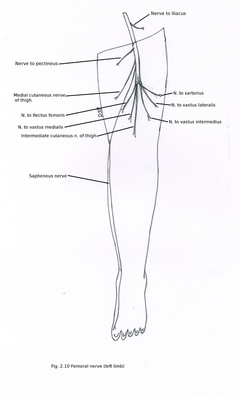
3. Femoral nerve and its branches :
It is a branch of lumbar plexus. Formed by dorsal branches of ventral rami of L2,L3 and L4. It appears near lateral border of psoas major after its formation. Goes down between psoas major and iliacus then deep to fascia iliaca. Then it goes deep to inguinal ligament into femoral triangle. In femoral triangle divides into anterior and posterior division. Lateral circumflex femoral artery lies in between two divisions.
Branches :
From trunk
1. Nerve to Iliacus muscle :
It supply iliacus inside abdomen.
2. Nerve to Pectineus :
It lies medial to femoral nerve and posterior to femoral sheath passes deep to inguinal ligament supply pectineus muscle from anterior surface.
3. Vascular branch :
To proximal part of femoral artery.
From anterior division
1. Intermediate femoral cutaneous nerve :
After piercing fascia lata 8 cm below inguinal ligament goes down in front of thigh supplying that area upto knee joint and also form patellar plexus.
2. Medial femoral cutaneous nerve :
It lies lateral to femoral artery but crosses artery near apex of femoral triangle from lateral to medial side. Divide into anterior and posterior branches. Anterior branch present in relation with anterior aspect of sartorius supply medial aspect of thigh upto medial side of knee and small infrapatellar branch connected with infrapatellar branch of saphenous nerve. Posterior branch runs alongwith posterior border of sartorius connected with saphenous nerve. Posterior branch joins with subsartorial plexus and supply skin of upper part of leg on medial side.
3. Nerve to Sartorius :
It may be separate branch of femoral or arise alongwith intermediate femoral cutaneous nerve.
From posterior division
1. Saphenous nerve :
It is longest cutaneous nerve. It is lateral to femoral artery in femoral triangle and upper part of adductor canal. In adductor canal it crosses femoral artery from lateral to medial side anteriorly. It gives a branch to form subsartorial plexus. Then it pierces fascia lata on postero medial aspect of knee in between tendons of sartorius and gracialis gives a cutaneous branch to prepatellar skin. Then it goes down alongwith great saphenous vein along medial border of tibia upto ankle. Supply skin on medial aspect of dorsum of foot upto first metatarso-phalangeal joint.
2. Muscular branches :
It supply quadriceps femoris. Branch to rectus femoris supply it from posterior aspect on proximal part and gives a branch to hip joint. Nerve to vastus lateralis forms a neurovascular bundle runs alongwith descending branch of lateral circumflex femoral artery and supply muscle giving a branch to knee joint. Nerve to vastus medialis goes downward in proximal part of adductor canal and supply muscle giving a branch to knee joint. Nerve to vastus intermedius by two or three branches anteriorly giving a branch to articularis genu and knee joint.
4. Deep inguinal group of lymph nodes :
These are three in number. Deep inguinal group of lymphnodes lies in relation with medial aspect of upper part of femoral vein draining lymph from vessels which runs alongwith branches of femoral vessels, glans penis in male, glans clitoris in female and from superficial inguinal group of lymph nodes.
5. Lateral femoral cutaneous nerve :
It is a branch of lumbar plexus. Dorsal branches of ventral rami L2, L3 form. It goes laterally from lateral border of psoas major towards anterior superior iliac spine. It divides into two branches anterior and posterior. Anterior branch supply anterior and lateral part of upper part of thigh upto knee. Posterior branch supply skin of greater trochanter upto middle part of thigh and also gluteal region.
6. Femoral branch of genitofemoral nerve :
It is a branch of lumbar plexus from L1. After its division from genitofemoral nerve enters inside lateral compartment of femoral sheath. Piercing femoral sheath anteriorly supply skin of femoral triangle.
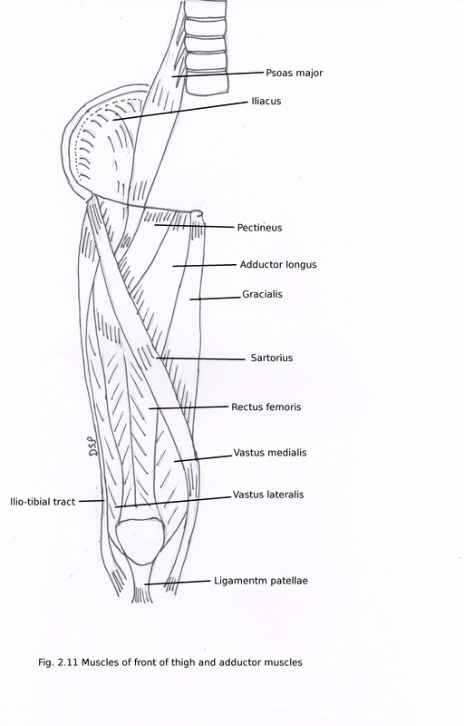
Muscles of front of thigh
These are sartorius, rectus femoris, vastus medialis, vastus intermedius, vastus lateralis, articularis genu.
Sartorius:
It is longest muscle.
Origin : It shows origin from anterior superior iliac spine and upper part of notch below it.
Insertion : It shows insertion on upper part of medial surface of tibia anterior to insertion of gracialis and semitendinosus. At insertion shape is like an inverted hockey stick.
Nerve supply : It receives nerve supply from anterior division of femoral nerve.
Action : Flexion, abduction, lateral rotation of hip joint.
Flexion, medial rotation of knee joint when it is in semiflexed position.
Quadriceps femoris
It is formed by extensor of knee joint. These are rectus femoris, vastus medialis, vastus intermedius and vastus lateralis.
A sesamoid bone patella is present in tendon of quadriceps femoris. Tendon shows attachment on tibial tuberosity.
Rectus femoris :
Origin : It shows origin from two heads straight and reflected. Straight head shows origin from anterior inferior iliac spine on its upper part. Reflected head shows origin from a groove in upper part of acetabulum and some fibres from capsule of hip joint.
It is a bipennate muscle. Two heads unite with each other at an acute angle and forms an aponeurosis. Aponeurosis forms a narrow flat tendon in lower part.
Insertion : It shows insertion of its tendon on base of patella.
Nerve supply : It receives nerve supply from posterior division of femoral nerve.
Action : Extension of knee joint and flexion of hip joint.
Vastus medialis :
Origin : It shows origin from a continues line from lower part of intertrochantric line, spiral line, medial lip of linea aspera, upper part of medial supracondylar line.
Insertion : It shows insertion on medial margin of patella in its upper two third part and base of patella in its medial one third part .
Nerve supply : It receives nerve supply from nerve to vastus medialis a branch of posterior division of femoral nerve.
Action : Extension of knee joint and prevent patella from lateral displacement.
Vastus lateralis :
Origin : It shows origin from a continues line from upper part of intertrochantric line, anterior and lower border of greater trochanter, lateral lip of gluteal tuberosity, proximal part of lateral lip of linea aspera, some fibres from lateral intermuscular septum.
Insertion : It shows insertion on lateral margin of patella in its upper one third part and base of patella in its lateral two third part .
Nerve supply : It receives nerve supply from posterior division of femoral nerve.
Action : Extension of knee joint.
Vastus intermedius :
Origin : It shows origin from anterior and lateral surface of upper two third part shaft of femur.
Insertion : It shows insertion on base of patella forming deep part of quadriceps femoris.
Nerve supply : It receives nerve supply from posterior division of femoral nerve.
Action : Extension of knee joint.
Articularis genu :
Origin : It shows origin by slips from anterior surface of lower part shaft of femur.
Insertion : It shows insertion on proximal reflexion of synovial membrane of knee joint.
Nerve supply : It receives nerve supply from nerve to vastus intermedius from posterior division of femoral nerve.
Action : pull upward synovial membrane.
Other muscles :
Iliacus :
Origin : It is a triangular muscle. It shows origin from upper two third part of iliac fossa, inner lip of iliac crest, ventral part of sacro-iliac ligament, ilio-lumbar ligament and ala of sacrum.
Insertion : It shows insertion on lesser trochanter of femur.
Nerve supply : It receives nerve supply from trunk of femoral nerve from L2,L3.
Action : Flexion of hip joint and lateral rotator of femur.
Psoas major :
Origin : It shows origin from anterior surface and transverse process of 5 lumbar vertebrae. It shows five digitations of origin. It goes down in relation to pelvic brim, deep to inguinal ligament forms a tendon.
Insertion : It shows insertion on anterior surface of lesser trochanter of femur.
Nerve supply : It receives nerve supply from lumbar plexus L1, L2 partly L3.
Action : Alongwith Iliacus flexion of hip joint.
Pectineus :
Origin : It shows origin from pecten pubis and from adjoining bone.
Insertion : It shows insertion on shaft of femur on posterior aspect in a line from lesser trochanter to linea aspera.
Nerve supply : It receives nerve supply from trunk of femoral nerve L2, L3 and obturator nerve or accessory obturator nerve (if present) L3.
Action : Adduction and flexion of hip joint.
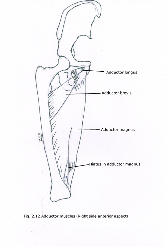
Adductor longus :
Origin : It shows origin as a tendon from anterior surface of pubis at an angle formed by pubic crest and pubic symphysis. Riders bone a sesamoid bone may present in tendon of adductor longus.
Insertion : It shows insertion on linea aspera of shaft of femur on posterior aspect in its middle third part.
Nerve supply : It receives nerve supply from anterior division of obturator nerve L2, L3, L4.
Action : Adduction and medial rotation of hip joint.
These are sartorius, rectus femoris, vastus medialis, vastus intermedius, vastus lateralis, articularis genu.
Sartorius:
It is longest muscle.
Origin : It shows origin from anterior superior iliac spine and upper part of notch below it.
Insertion : It shows insertion on upper part of medial surface of tibia anterior to insertion of gracialis and semitendinosus. At insertion shape is like an inverted hockey stick.
Nerve supply : It receives nerve supply from anterior division of femoral nerve.
Action : Flexion, abduction, lateral rotation of hip joint.
Flexion, medial rotation of knee joint when it is in semiflexed position.
Quadriceps femoris
It is formed by extensor of knee joint. These are rectus femoris, vastus medialis, vastus intermedius and vastus lateralis.
A sesamoid bone patella is present in tendon of quadriceps femoris. Tendon shows attachment on tibial tuberosity.
Rectus femoris :
Origin : It shows origin from two heads straight and reflected. Straight head shows origin from anterior inferior iliac spine on its upper part. Reflected head shows origin from a groove in upper part of acetabulum and some fibres from capsule of hip joint.
It is a bipennate muscle. Two heads unite with each other at an acute angle and forms an aponeurosis. Aponeurosis forms a narrow flat tendon in lower part.
Insertion : It shows insertion of its tendon on base of patella.
Nerve supply : It receives nerve supply from posterior division of femoral nerve.
Action : Extension of knee joint and flexion of hip joint.
Vastus medialis :
Origin : It shows origin from a continues line from lower part of intertrochantric line, spiral line, medial lip of linea aspera, upper part of medial supracondylar line.
Insertion : It shows insertion on medial margin of patella in its upper two third part and base of patella in its medial one third part .
Nerve supply : It receives nerve supply from nerve to vastus medialis a branch of posterior division of femoral nerve.
Action : Extension of knee joint and prevent patella from lateral displacement.
Vastus lateralis :
Origin : It shows origin from a continues line from upper part of intertrochantric line, anterior and lower border of greater trochanter, lateral lip of gluteal tuberosity, proximal part of lateral lip of linea aspera, some fibres from lateral intermuscular septum.
Insertion : It shows insertion on lateral margin of patella in its upper one third part and base of patella in its lateral two third part .
Nerve supply : It receives nerve supply from posterior division of femoral nerve.
Action : Extension of knee joint.
Vastus intermedius :
Origin : It shows origin from anterior and lateral surface of upper two third part shaft of femur.
Insertion : It shows insertion on base of patella forming deep part of quadriceps femoris.
Nerve supply : It receives nerve supply from posterior division of femoral nerve.
Action : Extension of knee joint.
Articularis genu :
Origin : It shows origin by slips from anterior surface of lower part shaft of femur.
Insertion : It shows insertion on proximal reflexion of synovial membrane of knee joint.
Nerve supply : It receives nerve supply from nerve to vastus intermedius from posterior division of femoral nerve.
Action : pull upward synovial membrane.
Other muscles :
Iliacus :
Origin : It is a triangular muscle. It shows origin from upper two third part of iliac fossa, inner lip of iliac crest, ventral part of sacro-iliac ligament, ilio-lumbar ligament and ala of sacrum.
Insertion : It shows insertion on lesser trochanter of femur.
Nerve supply : It receives nerve supply from trunk of femoral nerve from L2,L3.
Action : Flexion of hip joint and lateral rotator of femur.
Psoas major :
Origin : It shows origin from anterior surface and transverse process of 5 lumbar vertebrae. It shows five digitations of origin. It goes down in relation to pelvic brim, deep to inguinal ligament forms a tendon.
Insertion : It shows insertion on anterior surface of lesser trochanter of femur.
Nerve supply : It receives nerve supply from lumbar plexus L1, L2 partly L3.
Action : Alongwith Iliacus flexion of hip joint.
Pectineus :
Origin : It shows origin from pecten pubis and from adjoining bone.
Insertion : It shows insertion on shaft of femur on posterior aspect in a line from lesser trochanter to linea aspera.
Nerve supply : It receives nerve supply from trunk of femoral nerve L2, L3 and obturator nerve or accessory obturator nerve (if present) L3.
Action : Adduction and flexion of hip joint.
Adductor longus :
Origin : It shows origin as a tendon from anterior surface of pubis at an angle formed by pubic crest and pubic symphysis. Riders bone a sesamoid bone may present in tendon of adductor longus.
Insertion : It shows insertion on linea aspera of shaft of femur on posterior aspect in its middle third part.
Nerve supply : It receives nerve supply from anterior division of obturator nerve L2, L3, L4.
Action : Adduction and medial rotation of hip joint.
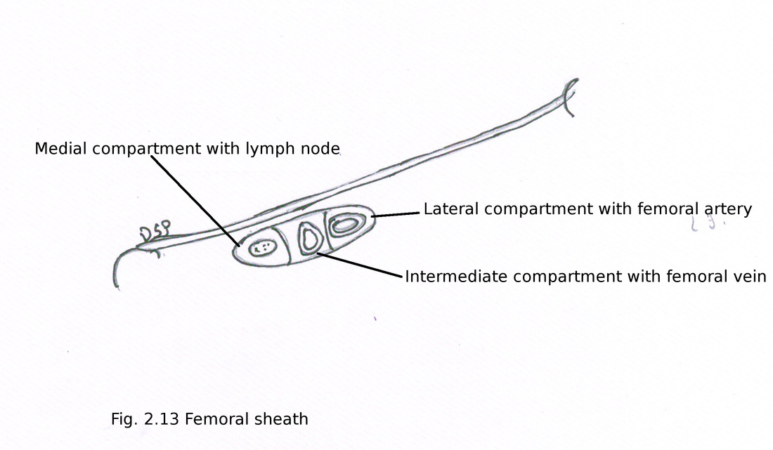
Femoral sheath
It is funnel like formed by fascia. Anteriorly formed by fascia transversalis and posteriorly by fascia iliaca. It covers femoral vessel about 3-4 cm below inguinal ligament. It help in free movement of vessels below inguinal ligament. At the time of birth it is short but elongation takes place after sometime when regular extension of hip starts. Lateral wall of it vertical and medial wall goes downward and laterally. Structures piercing are femoral branch of genitofemoral nerve, great saphenous vein, superficial epigastric artery, superficial circumflex iliac artery, superficial external pudendal artery.
Division: Two antero-posterior septa divide sheath into three compartments lateral, intermediate, medial. Lateral compartment contains femoral artery and femoral branch of genitofemoral nerve, intermediate compartment contains femoral vein, medial compartment known as femoral canal contains lymph vessels and a lymphnode. Femoral canal is about 1.25 cm in length. Base is formed by femoral ring. Femoral ring anteriorly shows inguinal ligament and posteriorly shows pectineus muscle and fascia, medially shows base of lacunar ligament, laterally shows femoral vein. Femoral ring is larger in female due to wider pelvis. Femoral septum closes femoral ring.
Applied : Femoral hernia a peritoneal pouch goes downward through femoral ring and then into femoral canal finally goes anteriorly through saphenous opening then upwards.
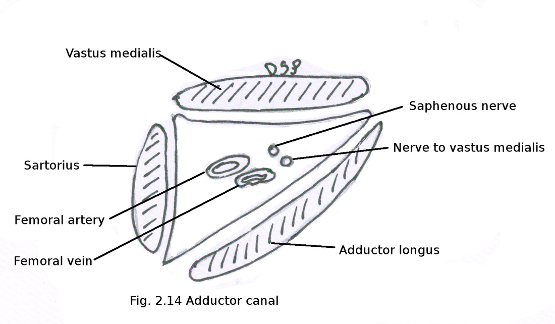
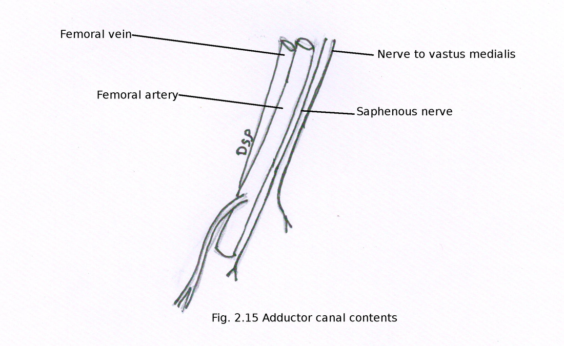
Adductor canal (Hunter's canal; Subsartorial canal)
It is an intermuscular canal deep to sartorius on medial aspect of thigh in middle one third part of thigh. It starts from apex of femoral triangle to lower attachment of adductor magnus. It is triangular in section.
Boundaries :
Anterolaterally : vastus medialis
Posteromedially : adductor longus above, adductor magnus below
Roof : covered by strong fascia which extends between two margins. Sartorius muscle present over this fascia. Therefore known as subsartorial fascia. Nerve plexus (subsartorial plexus) present in this fascia formed by branches from saphenous nerve, posterior branch of medial femoral cutaneous nerve, anterior division of obturator nerve.
Contents :
Femoral artery, femoral vein, descending genicular artery (branch of femoral), muscular branches of femoral artery, saphenous nerve, nerve to vastus medialis, terminal part of profunda femoris artery and sometimes posterior division of obturator nerve.
Femoral artery enters inside canal from apex of femoral triangle and exit is at fifth opening in adductor magnus.
Femoral vein present on posterior aspect of femoral artery above but it lies posterolaterally below.
Saphenous nerve crosses femoral artery from lateral to medial side from upper to lower part of canal.
Adductor region
It present on medial aspect of thigh. It is in between medial intermuscular septum and posterior intermuscular septum.
Contents :
Muscles : Gracialis, pectineus, adductor longus, obturator externus, adductor brevis, adductor magnus
Nerves : Obturator nerve
Artery : Obturator artery and profunda femoris artery.
Gracialis :
Origin : It shows origin on anterior surface of lower part of body of pubis, inferior ramus of pubis and ramus of ischium.
Insertion : It shows insertion below medial condyle of tibia by a tendon in upper part of medial surface of tibia. Insertion lies in between insertion of sartorius anteriorly and semitendinosus posteriorly.
Nerve supply : It receives nerve supply from anterior division of obturator nerve L2, L3.
Action : Adduction of hip joint, medial rotation of leg at knee joint when knee is semiflexed, rotate femur with pelvis laterally when foot is on ground.
Obturator externus :
Origin : It shows origin from on anterior surface of obturator membrane alongwith medial margin of obturator foramen and part of pubis, ischial ramus around it except upper part near obturator notch.
Insertion : It forms a tendon which goes backward, laterally and upwards on the back of neck of femur. It shows insertion on trochantric fossa of femur.
Nerve supply : It receives nerve supply from posterior division of obturator nerve L3, L4.
Action : It is a postural muscle. Lateral rotator of hip joint.
Adductor brevis :
It is triangular in shape.
Origin : It shows origin from anterior surface of body of pubis and inferior ramus of pubis between origin of gracialis and obturator externus.
Insertion : It shows insertion on a line from lesser trochanter to linea aspera of shaft of femur on posterior aspect in its upper part.
Nerve supply : It receives nerve supply from anterior division of obturator nerve L2, L3.
Action : Adduction of hip joint.
Adductor magnus :
It is a large triangular muscle.
Origin : It shows origin from inferior ramus of pubis, ischiopubic ramus and inferolateral part of ischial tuberosity.
Insertion : It shows insertion of pubic fibres on gluteal tuberosity in its medial margin, ischiopubic fibres (adductor component) on medial margin of linea aspera shaft of femur on posterior and upper part of medial supracondylar line, fibres from ischial tuberosity (hamstring component) on adductor tubercle.
Nerve supply : It receives nerve supply adductor component from obturator nerve and hamstring component from tibial division of sciatic nerve L2, L3, L4.
Action : Adduction and medial rotation at hip joint.
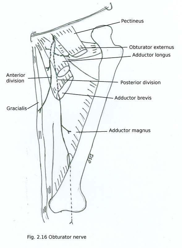
Obturator nerve :
It is a branch from lumbar plexus. Formed by ventral branches of ventral rami of L2,L3,L4.
Course : After its formation in the substance of psoas major muscle runs along medial border of it. It goes downward crossing ala of sacrum posterior to common iliac vessels. It runs on lateral wall of pelvis by crossing pelvic brim. Enters obturator canal with obturator vessels. In female it is present on lateral wall of ovarian fossa. Enters in adductor compartment and divides into two branches anterior and posterior.
Anterior branch : It passes anterior to obturator externus and adductor brevis; posterior to petineus and adductor longus. It goes down near lower margin of adductor longus joins with medial cutaneous nerve and saphenous nerve (branches of femoral nerve) and forms subsartorial plexus to supply medial side of thigh. It also supply femoral artery, branch to hip joint near obturator canal. Muscular branches are given to adductor longus, gracialis, adductor brevis, pectineus.
Posterior branch : It goes posterior by piercing obturator externus. Goes downward posterior to adductor brevis and anterior to adductor magnus. Muscular branches are given to obturator externus, adductor part of adductor magnus and adductor brevis (if no branch from anterior division). It gives a branch to knee joint. It goes down through opening in adductor magnus alongwith femoral vessels in popliteal fossa supply capsule of knee joint, popliteal artery, cruciate ligaments.
Applied anatomy :
1. Referred pain to hip, knee and medial aspect of thigh in disease of ovary.
2. Nerve entrapment syndrome is a condition in which chronic pain on medial side of thigh due to big adductor muscles common in athletes.
Accessory obturator nerve
It a small branch present sometimes. It is a small branch from ventral branches ventral rami of L3, L4. Goes down along medial margin of psoas major after crossing superior pubic ramus goes posterior to pectineus and gives out branches. Branches are given to pectineus, hip joint, a branch joins with anterior division of obturator nerve.
Obturator artery :
It is a branch of anterior division of internal iliac artery. It goes downward along lateral pelvic wall upto obturator foramen. It lies over fascia of obturator internus. Ureter crosses it alonwith vas deferens in male and in female ovary lies medial to it. Runs alonwith obturator nerve and vein. In pelvis branches are given to iliac bone, iliacus, anastomosing branch to iliolumbar artery, vesical branch sometimes, pubic branch. Comes out through obturator canal in adductor region. Here it divide into anterior and posterior branches. Anterior branch goes down on obturator membrane supply obturator externus, pectineus, gracialis, adductors of thigh. Finally anterior branch anastomose with posterior branch and medial circumflex femoral artery. Posterior branch goes downward along posterior line of obturator foramen finally anastomose with anterior division. It gives a branch to hip joint.
Sometimes pubic branch of inferior epigastric artery replace obturator artery. It is present on lateral aspect of femoral ring. This may get ruptured during surgery of femoral and inguinal hernia while dilating femoral ring.
