PERICARDIUM AND HEART
Pericardium
It is a fibroserous sac present around heart and great vessels. It is located in middle mediastinum. Parts of pericardium are 1) fibrous pericardium and 2) serous pericardium
Pericardium
It is a fibroserous sac present around heart and great vessels. It is located in middle mediastinum. Parts of pericardium are 1) fibrous pericardium and 2) serous pericardium
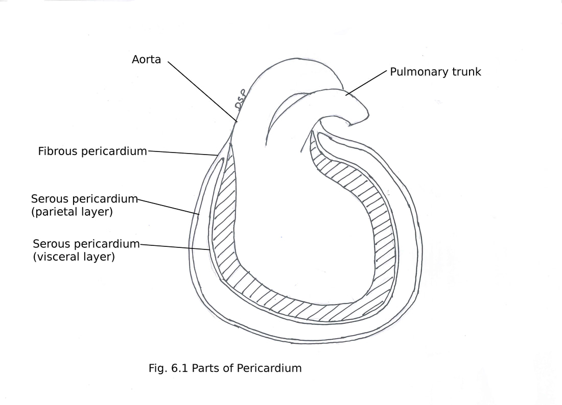
1) fibrous pericardium :
It shows a cone shaped sac. It is made up of strong fibrous tissue.
Attachments : Above : It shows attachment on adventitia of ascending aorta and pulmonary trunk and then continue with the pre-tracheal layer of deep cervical fascia.
Anteriorly : It shows attachment on posterior surface of sternum by superior and inferior sternopericardial ligament.
Below : It shows attachment with the upper surface of central tendon and a part of the left
diaphragm.
Relations :
In front : It comes in relation with anterior thoracic wall separated by anterior margins of right and left lungs and pleurae. Below the left 4th and 5th costal cartilage it comes in direct relation with anterior thoracic wall.
Posteriorly : It comes in relation with right and left principal bronchi, posterior part of mediastinal aspect of right and left lungs, the oesophagus, oesophageal plexus, descending thoracic aorta, thoracic duct, azygos and hemi-azygos veins.
On right and left sides : It comes in relation with mediastinal pleura, mediastinal surface of lung, phrenic nerves and pericardiophrenic vessels.
Below : It comes in relation with left lobe of liver and fundus of stomach separated by diaphragm.
Structures Piercing
Following structures pierce it and receives extension from it which shows attachment on these structures ascending aorta, right and left pulmonary arteries,superior and inferior vena cava and four pulmonary veins.
2) Serous pericardium
It is a closed serous sac present inside fibrous pericardium. It shows two layers outer parietal pericardium and inner visceral pericardium or epicardium. Parietal layer present on inner aspect of fibrous pericardium firmly attached with it. Visceral layer lines heart and great vessels firmly attached with it. The two layers shows continuation with each other at the roots of the great vessels after reflexion. The pericardial cavity is formed between the parietal pericardium and the visceral pericardium. It contains serous fluid forming a thin film.
Contents of the Pericardium : Following are contents of pericardium heart, cardiac vessels, cardiac nerves, pulmonary trunk, ascending aorta, superior vena cava (lower part), inferior vena cava (terminal part ) and part of the pulmonary veins (terminal part ) .
The parietal and visceral pericardium forms reflexions at the roots of the great vessels is arranged in form of two tubes arterial tube enclosing the ascending aorta and the pulmonary trunk (arterial end of the heart tube) and the venous tube enclosing the venae cavae and pulmonary veins (venous end of the heart tube).
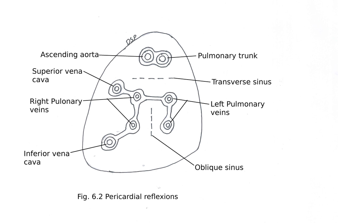
Sinuses of pericardium
Transverse sinus : It is a horizontal gap between two arterial and venous ends of serous pericardium and covered by visceral layer.
Boundaries : Anteriorly shows ascending aorta and pulmonary trunk, posteriorly shows superior vena cava and left atrium.
Oblique sinus : It is a small gap behind the left atrium. It is open below. It is in between parietal and visceral layers.
Boundaries : Anteriorly shows left atrium, posteriorly parietal pericardium, on right side shows two right pulmonary veins with inferior vena cava and on left side shows left two pulmonary veins.
Fold of left vena cava (Fold of Marshall) : It is a fold of serous pericardium from left pulmonary artery to the upper left pulmonary vein. It is formed by a fibrous ligament which is a remnant
of the obliterated left common cardinal vein (left duct of Cuvier). It is continuous above with terminal part of left superior intercostal vein and below continue as oblique vein of left atrium.
Arterial Supply : It receives blood supply from branches of internal thoracic artery, musculophrenic and descending thoracic aorta.
Venous drainage : It drains into azygos veins.
Nerve supply : Visceral layer receives nerve supply from vagus nerve and sympathetic nerves (insensitive to pain), fibrous and parietal layer from phrenic nerves (sensitive to pain).
Applied Anatomy :
1) Pericardial effusion : It is collection of fluid in the pericardial cavity.
2) Aspiration of fluid can be done by a needle puncturing 4th or 5th intercostal space close to lateral margin of sternum or angle between xiphoid process and left costal margin needle directed upwards backwards.
3) Cardiac tamponade : Fluid accumulation in pericardial sac decreases diastolic capacity of heart because of pressure of fluid so it decreases decreased cardiac output known as cardiac tamponade.
HEART
The heart is a hollow muscular organ present in the middle mediastinum. It is conical in shape. It is covered by pericardium. It shows four chambers right and left atria and right and left ventricles.
External Features
It is 12 cm from base to apex and 8 cm in transverse diameter and weighs about 300 g in males and 250 g in females. Atria present above and behind the ventricles. They shows grooves which separates atria from ventricles by an atrioventricular groove, atria are separated by an inter-atrial groove and between ventricles shows interventricular groove
Parts of heart
Heart shows an apex, base, surfaces sternocostal (anterior), diaphragmatic (inferior) and right and left (pulmonary) surfaces, four borders right, inferior, left and upper.
Apex is conical part formed by the left ventricle directed downwards, forwards and to the left. Apex lies in 5th intercostal space medial to left midclavicular line.
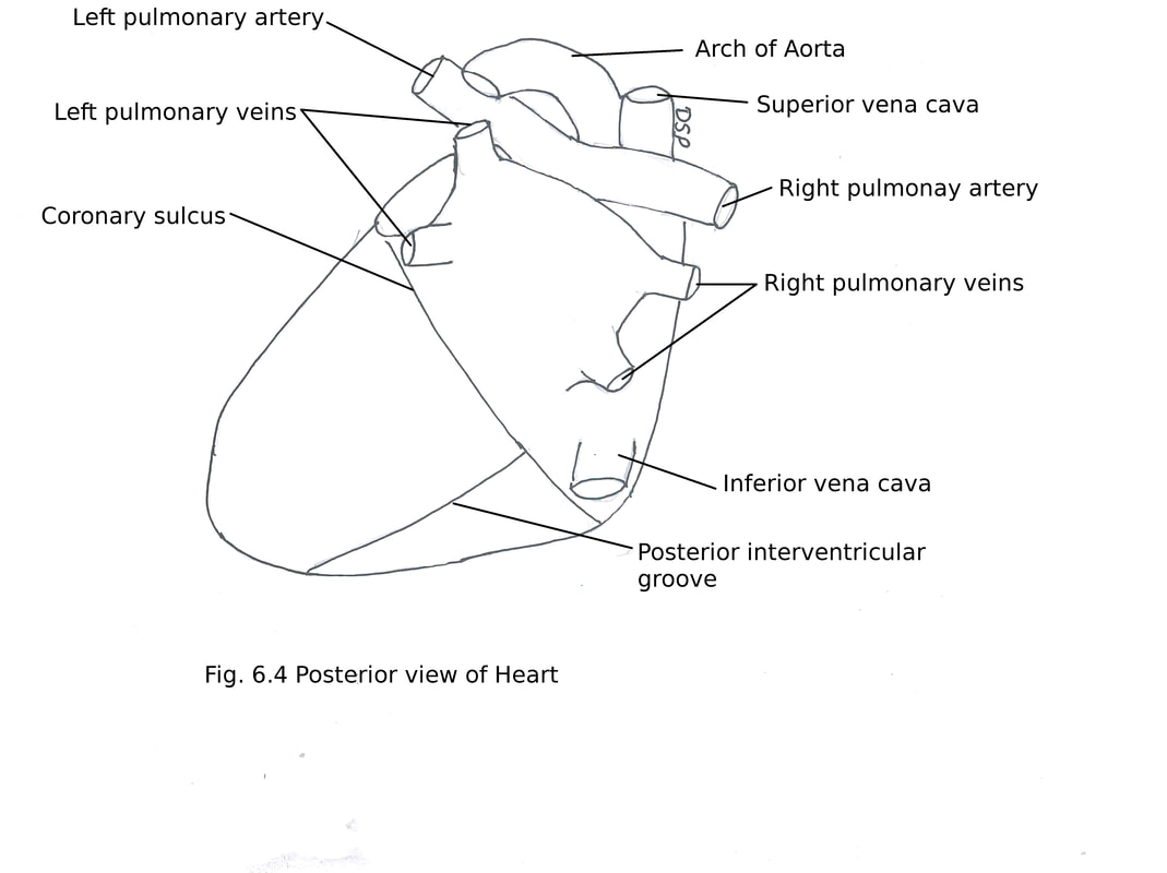
The base of the heart is formed by posterior surface. It is formed by left atrium and part of right atrium. It extends above up to bifurcation of pulmonary trunk, below to posterior part of the atrioventricular groove which contains coronary sinus and anastomosis of right and left coronary arteries. Right and left side limited by right border of the right atrium and left border of left atrium. Superior and inferior vena cava opens in the upper and lower part of right atrium. Four pulmonary veins shows openings on posterior surface of left atrium.
Right border is formed by the right atrium. It is slightly convex and extends from opening of superior vena cava to inferior vena cava on its right side. It separates base of heart from sternocostal surface.
Left border is formed by the left ventricle and part of left auricle. It is round and separates sternocostal surface and left surfaces.
Inferior border is horizontal and formed by right ventricle and near the apex formed by part of left ventricle. It separates sternocostal surface from diaphragmatic surface.
Upper border formed by left atrium. Superior vena cava enters in right atrium in its upper most part.
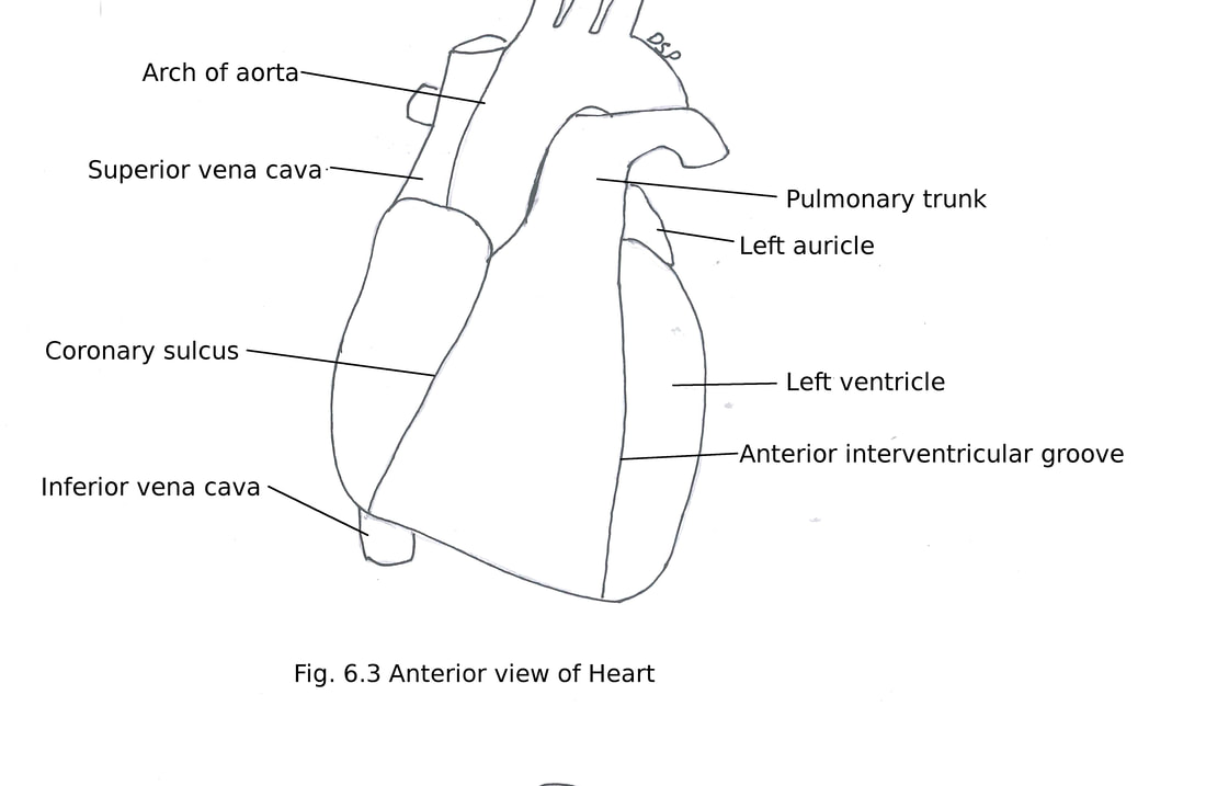
Sternocostal (anterior) surface faces anteriorly and upwards. It is formed by anterior surface of right atrium and right ventricle (⅔), part of anterior surface of left ventricle (⅓) and left auricle.
Anterior part of atrioventricular groove is seen going downwards and towards right. It shows right coronary artery running in this groove. Anterior interventricular groove present on this surface indicates interventricular septum lies parallel to the left border of the heart. Anterior interventricular groove shows anterior interventricular branch of left coronary artery and great cardiac vein.
This surface is shows covering of pericardium. It comes in relation with posterior surface of the body of sternum, sternocostalis muscle, 3rd to 6th costal cartilages of right and left side. It lies more on left side because of position of heart. It is covered by the lungs and pleura.
Diaphragmatic (inferior) surface faces downward, backwards. It is formed mainly by left ventricle with partly contribution from right ventricle. It rests on central tendon of diaphragm. It shows posterior interventricular groove. This groove contains posterior interventricular branch of the right
coronary artery, middle cardiac vein and anastomosis of interventricular branches of right and left coronary arteries.
Left (pulmonary) surface is formed mainly by left ventricle and partly by left atrium with left auricle. It faces upward, backward and towards left. It shows left part of the atrioventricular groove. This groove contains trunk and circumflex branch of left coronary artery, terminal part of great cardiac vein, starting point of coronary sinus. It comes in relation with left phrenic nerve, left pericardiophrenic vessels separated by pericardium.
Right surface is formed by wall of right atrium. Sulcus terminalis is visible here lies at junction of sternocostal and right surfaces.
Grooves or Sulci
Circular atrioventricular groove present in between atria and ventricles. It shows trunks of coronary arteries in it. Its upper part is crossed anteriorly by the ascending aorta and pulmonary trunk. Interatrial groove present in between two atria seen on posterior aspect. Anterior interventricular groove is present on sternocostal surface lie near to the left margin of the heart. Posterior interventricular groove is present on the diaphragmatic (inferior) surface of heart. Anterior interventricular grooves continue as a posterior interventricular groove near apex of heart.
Circulation of blood in heart :
Right atrium receives deoxygenated blood from superior and inferior venae cava (body) and coronary sinus. Then it pumps blood through the right atrioventricular orifice (guarded by tricuspid valve) to the right ventricle. Right ventricle pumps blood to pulmonary trunk which will go to lungs. Oxygenated blood from lung comes to left atrium via 4 pulmonary veins. Left atrium pumps blood to left ventricle through the left atrioventricular orifice (guarded by mitral valve). Finally left ventricle pump blood into the aortic sinuses and ascending aorta.
Heart shows an apex, base, surfaces sternocostal (anterior), diaphragmatic (inferior) and right and left (pulmonary) surfaces, four borders right, inferior, left and upper.
Apex is conical part formed by the left ventricle directed downwards, forwards and to the left. Apex lies in 5th intercostal space medial to left midclavicular line.
The base of the heart is formed by posterior surface. It is formed by left atrium and part of right atrium. It extends above up to bifurcation of pulmonary trunk, below to posterior part of the atrioventricular groove which contains coronary sinus and anastomosis of right and left coronary arteries. Right and left side limited by right border of the right atrium and left border of left atrium. Superior and inferior vena cava opens in the upper and lower part of right atrium. Four pulmonary veins shows openings on posterior surface of left atrium.
Right border is formed by the right atrium. It is slightly convex and extends from opening of superior vena cava to inferior vena cava on its right side. It separates base of heart from sternocostal surface.
Left border is formed by the left ventricle and part of left auricle. It is round and separates sternocostal surface and left surfaces.
Inferior border is horizontal and formed by right ventricle and near the apex formed by part of left ventricle. It separates sternocostal surface from diaphragmatic surface.
Upper border formed by left atrium. Superior vena cava enters in right atrium in its upper most part.
Sternocostal (anterior) surface faces anteriorly and upwards. It is formed by anterior surface of right atrium and right ventricle (⅔), part of anterior surface of left ventricle (⅓) and left auricle.
Anterior part of atrioventricular groove is seen going downwards and towards right. It shows right coronary artery running in this groove. Anterior interventricular groove present on this surface indicates interventricular septum lies parallel to the left border of the heart. Anterior interventricular groove shows anterior interventricular branch of left coronary artery and great cardiac vein.
This surface is shows covering of pericardium. It comes in relation with posterior surface of the body of sternum, sternocostalis muscle, 3rd to 6th costal cartilages of right and left side. It lies more on left side because of position of heart. It is covered by the lungs and pleura.
Diaphragmatic (inferior) surface faces downward, backwards. It is formed mainly by left ventricle with partly contribution from right ventricle. It rests on central tendon of diaphragm. It shows posterior interventricular groove. This groove contains posterior interventricular branch of the right
coronary artery, middle cardiac vein and anastomosis of interventricular branches of right and left coronary arteries.
Left (pulmonary) surface is formed mainly by left ventricle and partly by left atrium with left auricle. It faces upward, backward and towards left. It shows left part of the atrioventricular groove. This groove contains trunk and circumflex branch of left coronary artery, terminal part of great cardiac vein, starting point of coronary sinus. It comes in relation with left phrenic nerve, left pericardiophrenic vessels separated by pericardium.
Right surface is formed by wall of right atrium. Sulcus terminalis is visible here lies at junction of sternocostal and right surfaces.
Grooves or Sulci
Circular atrioventricular groove present in between atria and ventricles. It shows trunks of coronary arteries in it. Its upper part is crossed anteriorly by the ascending aorta and pulmonary trunk. Interatrial groove present in between two atria seen on posterior aspect. Anterior interventricular groove is present on sternocostal surface lie near to the left margin of the heart. Posterior interventricular groove is present on the diaphragmatic (inferior) surface of heart. Anterior interventricular grooves continue as a posterior interventricular groove near apex of heart.
Circulation of blood in heart :
Right atrium receives deoxygenated blood from superior and inferior venae cava (body) and coronary sinus. Then it pumps blood through the right atrioventricular orifice (guarded by tricuspid valve) to the right ventricle. Right ventricle pumps blood to pulmonary trunk which will go to lungs. Oxygenated blood from lung comes to left atrium via 4 pulmonary veins. Left atrium pumps blood to left ventricle through the left atrioventricular orifice (guarded by mitral valve). Finally left ventricle pump blood into the aortic sinuses and ascending aorta.
Chambers of heart
Right atrium
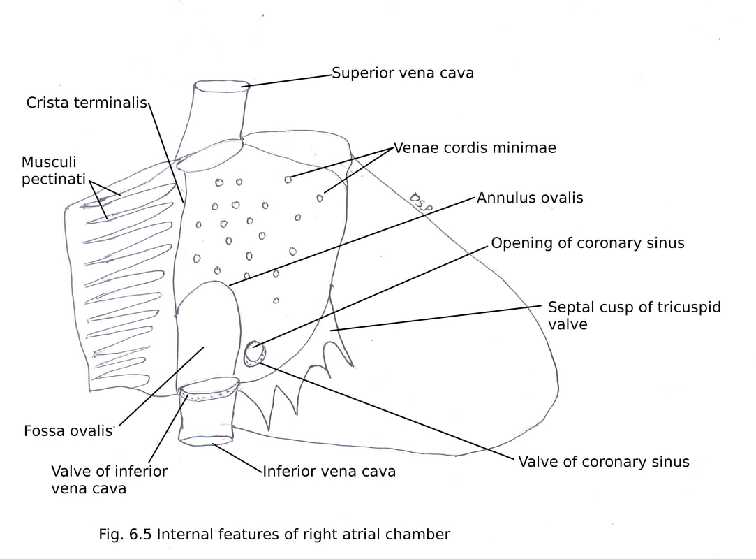
It receives deoxygenated blood from body. Then pumps to right ventricle through right atrioventricular opening guarded by tricuspid valve. Its walls forms right surface (pulmonary surface), right upper sternocostal surface, right side of base and parts of the upper border. Superior vena cava opens in it at upper part and inferior vena cava at lower posterior part. A projection at upper end and to left is present forming right auricle. Along right border of atrium externally a shallow vertical groove sulcus terminalis is present which passes from superior to inferior vena cava. Internally it corresponds to crista terminalis. Sinuatrial (SA) node is present in upper part of this sulcus. Right atrioventricular groove containing right coronary artery and small cardiac vein which separates right atrium from the right ventricle. It receives following vessels superior vena cava, inferior vena cava, coronary sinus, anterior cardiac veins, venae cordis minimi and right marginal vein.
Internal features of the right atrium can be divided into the following three parts (1) The Smooth posterior part or Sinus Venarum (2) Rough Anterior Part or Pectinate part including the auricle and (3) Septal Wall.
The Smooth posterior part or Sinus Venarum. It receives openings of the superior vena cava at the upper end, inferior vena cava at the lower end guarded by valve (Eustachian valve) and coronary sinus (guarded by Thebesian valve) between opening of inferior cava and the right atrioventricular orifice. Venae cordis minimi are numerous small veins draining minimal atrial veins from atrial walls. The intervenous tubercle small muscular projection below opening of superior vena cava. It regulate superior caval blood flow. Rough anterior part or pectinate part including the auricle presents transverse muscular ridges called musculi pectinati arises from crista terminalis. Crista terminalis is a smooth muscle ridge extends from upper part of atrial septum passes anterior to opening of superior vena cava. Below crista joins with right horn of valve of inferior vena cava. Septal wall shows fossa ovalis, above and to left of opening of inferior vena cava. Annulus ovalis or limbus fossa ovalis is a sharp margin of the fossa ovalis. Triangle of Koch is a triangular area bounded by septal leaflet of tricuspid valve, anteromedial margin of the opening of coronary sinus and tendon of Todaro (subendocardial ridge from the central fibrous body to left horn of the valve of inferior vena cava). A-V node lies here. Torus aorticus is an bulging in anterosuperior part of septum formed by right posterior aortic sinus.
Right atrium
It receives deoxygenated blood from body. Then pumps to right ventricle through right atrioventricular opening guarded by tricuspid valve. Its walls forms right surface (pulmonary surface), right upper sternocostal surface, right side of base and parts of the upper border. Superior vena cava opens in it at upper part and inferior vena cava at lower posterior part. A projection at upper end and to left is present forming right auricle. Along right border of atrium externally a shallow vertical groove sulcus terminalis is present which passes from superior to inferior vena cava. Internally it corresponds to crista terminalis. Sinuatrial (SA) node is present in upper part of this sulcus. Right atrioventricular groove containing right coronary artery and small cardiac vein which separates right atrium from the right ventricle. It receives following vessels superior vena cava, inferior vena cava, coronary sinus, anterior cardiac veins, venae cordis minimi and right marginal vein.
Internal features of the right atrium can be divided into the following three parts (1) The Smooth posterior part or Sinus Venarum (2) Rough Anterior Part or Pectinate part including the auricle and (3) Septal Wall.
The Smooth posterior part or Sinus Venarum. It receives openings of the superior vena cava at the upper end, inferior vena cava at the lower end guarded by valve (Eustachian valve) and coronary sinus (guarded by Thebesian valve) between opening of inferior cava and the right atrioventricular orifice. Venae cordis minimi are numerous small veins draining minimal atrial veins from atrial walls. The intervenous tubercle small muscular projection below opening of superior vena cava. It regulate superior caval blood flow. Rough anterior part or pectinate part including the auricle presents transverse muscular ridges called musculi pectinati arises from crista terminalis. Crista terminalis is a smooth muscle ridge extends from upper part of atrial septum passes anterior to opening of superior vena cava. Below crista joins with right horn of valve of inferior vena cava. Septal wall shows fossa ovalis, above and to left of opening of inferior vena cava. Annulus ovalis or limbus fossa ovalis is a sharp margin of the fossa ovalis. Triangle of Koch is a triangular area bounded by septal leaflet of tricuspid valve, anteromedial margin of the opening of coronary sinus and tendon of Todaro (subendocardial ridge from the central fibrous body to left horn of the valve of inferior vena cava). A-V node lies here. Torus aorticus is an bulging in anterosuperior part of septum formed by right posterior aortic sinus.

Right ventricle
Right ventricle forms sternocostal surface, part of the diaphragmatic surface and inferior border of heart. It shows two surfaces anterior (sternocostal) and inferior (diaphragmatic). Interior of right ventricle is semilunar and musculature is thinner as compared to left ventricle. Interior shows two parts (1) Inflowing rough part and (2) Outflowing smooth part. Inflowing part shows muscular
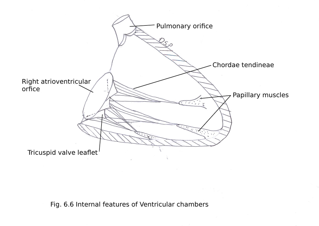
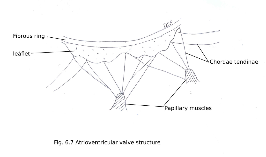
ridges known as trabeculae carneae. Supraventricular crest (muscular ridge) intervenes between inflowing and outflowing part. Inflowing part shows two orifices (1) Right atrioventricular (tricuspid orifice) tricuspid valve present here and (2) Pulmonary orifice. There are three cusps in the tricuspid valve named anterior, posterior and septal. Inflowing part in its inner aspect shows muscular ridges (trabeculae carneae) of three types ridges (fixed elevated), bridges (with two ends fixed free in middle) and pillars known as papillary muscles. Papillary muscles shows one end attached on ventricular wall and the other end attached on cusps of the tricuspid valve by chordae tendineae. Cusps of valves are three in number known as anterior, posterior and septal. Chordae tendineae supports cusps of atrioventricular valves formed by fibrocollagenous material. Three papillary muscles present in right ventricle anterior, posterior and septal. Septomarginal trabeculae or moderator band is a muscular ridge extending from the ventricular septum to the base of the anterior papillary muscle. Right branch of the AV bundle present in it.
Outflow part apex shows pulmonary orifice guarded by three semilunar cusps two in front and one behind. Margins of cusps shows a fibrous nodule at the center of margin and two thin lunulae at other side. cusp is formed by fold of endocardium. Above each cusp pulmonary trunk presents a dilatation known as pulmonary sinus.
The left atrium
It is a cuboidal chamber present posterior to right atrium. Anterosuperiorly shows a conical projection left auricle. It forms ⅔ part of base of heart. It shows four opening of pulmonary veins which receives oxygenated blood from lungs. It is communicated with left ventricle through the left atrioventricular (bicuspid or mitral) orifice guarded by bicuspid or mitral valve. Interior of left atrium is smooth. Some musculi pectinati is present inside. Some minimal cardiac veins opens directly in cavity from myocardium. Septal wall shows fossa lunata bounded by crescentic ridge concave upwards corresponds to fossa ovalis of right atrium.
Left ventricle
The left ventricle is conical in shape. Its wall is three times thicker as compared to right ventricle. It shows three surfaces sternocostal or anterior, diaphragmatic or inferior and left. It forms the apex of the heart, ⅓ part of the sternocostal surface, left border, left surface and the left 2/3 of the diaphragmatic surface. Interior of left ventricle shows inflow tract and outflow tract.
Inflow tract consists of left atrioventricular orifice guarded by bicuspid mitral valve and trabeculae carneae. Mitral valve also known as bicuspid valve shows two leaflets anterior and posterior. It conducts blood from the left atrium to left ventricle. There are two papillary muscles anterolateral and posteromedial. Chordae tendineae from both muscles shows attachment on cusps of the mitral valve.
The outflow tract leads to aortic opening. Which carry blood from left ventricle to ascending aorta. It is guarded by three semilunar cusps one in front and two behind. Margins of cusps shows a fibrous nodule at the center of free margin and two thin lunulae at other side. Cusp is formed by fold of endocardium. Above each cusp ascending aorta shows a dilatation known as aortic sinus of valsalva. Anterior aortic sinus shows origin of right coronary artery and the left posterior aortic sinus shows origin of left coronary artery.
Right ventricle forms sternocostal surface, part of the diaphragmatic surface and inferior border of heart. It shows two surfaces anterior (sternocostal) and inferior (diaphragmatic). Interior of right ventricle is semilunar and musculature is thinner as compared to left ventricle. Interior shows two parts (1) Inflowing rough part and (2) Outflowing smooth part. Inflowing part shows muscular
ridges known as trabeculae carneae. Supraventricular crest (muscular ridge) intervenes between inflowing and outflowing part. Inflowing part shows two orifices (1) Right atrioventricular (tricuspid orifice) tricuspid valve present here and (2) Pulmonary orifice. There are three cusps in the tricuspid valve named anterior, posterior and septal. Inflowing part in its inner aspect shows muscular ridges (trabeculae carneae) of three types ridges (fixed elevated), bridges (with two ends fixed free in middle) and pillars known as papillary muscles. Papillary muscles shows one end attached on ventricular wall and the other end attached on cusps of the tricuspid valve by chordae tendineae. Cusps of valves are three in number known as anterior, posterior and septal. Chordae tendineae supports cusps of atrioventricular valves formed by fibrocollagenous material. Three papillary muscles present in right ventricle anterior, posterior and septal. Septomarginal trabeculae or moderator band is a muscular ridge extending from the ventricular septum to the base of the anterior papillary muscle. Right branch of the AV bundle present in it.
Outflow part apex shows pulmonary orifice guarded by three semilunar cusps two in front and one behind. Margins of cusps shows a fibrous nodule at the center of margin and two thin lunulae at other side. cusp is formed by fold of endocardium. Above each cusp pulmonary trunk presents a dilatation known as pulmonary sinus.
The left atrium
It is a cuboidal chamber present posterior to right atrium. Anterosuperiorly shows a conical projection left auricle. It forms ⅔ part of base of heart. It shows four opening of pulmonary veins which receives oxygenated blood from lungs. It is communicated with left ventricle through the left atrioventricular (bicuspid or mitral) orifice guarded by bicuspid or mitral valve. Interior of left atrium is smooth. Some musculi pectinati is present inside. Some minimal cardiac veins opens directly in cavity from myocardium. Septal wall shows fossa lunata bounded by crescentic ridge concave upwards corresponds to fossa ovalis of right atrium.
Left ventricle
The left ventricle is conical in shape. Its wall is three times thicker as compared to right ventricle. It shows three surfaces sternocostal or anterior, diaphragmatic or inferior and left. It forms the apex of the heart, ⅓ part of the sternocostal surface, left border, left surface and the left 2/3 of the diaphragmatic surface. Interior of left ventricle shows inflow tract and outflow tract.
Inflow tract consists of left atrioventricular orifice guarded by bicuspid mitral valve and trabeculae carneae. Mitral valve also known as bicuspid valve shows two leaflets anterior and posterior. It conducts blood from the left atrium to left ventricle. There are two papillary muscles anterolateral and posteromedial. Chordae tendineae from both muscles shows attachment on cusps of the mitral valve.
The outflow tract leads to aortic opening. Which carry blood from left ventricle to ascending aorta. It is guarded by three semilunar cusps one in front and two behind. Margins of cusps shows a fibrous nodule at the center of free margin and two thin lunulae at other side. Cusp is formed by fold of endocardium. Above each cusp ascending aorta shows a dilatation known as aortic sinus of valsalva. Anterior aortic sinus shows origin of right coronary artery and the left posterior aortic sinus shows origin of left coronary artery.
Conducting system of heart
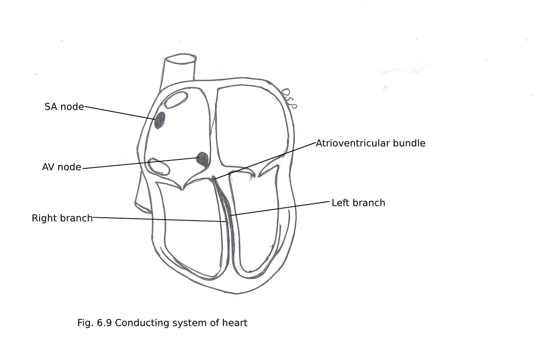
It shows specialized myocytes which can initiate and conduct impulse. It shows following parts : 1. Sinuatrial node (S. A. node), 2. Atrioventricular node (A. V. node), 3. Atrioventricular bundle (A. V. bundle or bundle of His) its two limb branches, 5. Right branch, 6. Left branch and 7. Purkinje fibres. The cardiac impulse begins at the S. A. node stimulates atrial musculature then reaches to A. V. node then it passes through A. V. bundle and through right and left limbs and purkinje fibres goes to ventricular wall.
1. Sinuatrial node (S. A. node) : It is elliptical structure. “P” (pacemaker cells) cells are prominent in this. It lies at a groove between right auricle and right side of superior vena the cava at upper part sulcus terminalis. It initiate cardiac impulse. To maintain heart rate at 70 per minute. It is also known as pacemaker.
2. Atrioventricular node (A. V. node) : It lies at right side of atrial septum above opening of coronary sinus. It is situated in triangle of Koch (bounded above by tendon of Todaro, below by the opening of coronary sinus and in front by base of the septal leaflet of tricuspid valve). It can generate impulse at 60 per minute.
3. Atrioventricular bundle (A. V. bundle or bundle of His) :
It arises from AV node. It traverses central fibrous body. It goes downwards after crossing AV ring reaches on dorsal margin of membranous part of ventricular septum. Here it divides into two branches right and left.
5. Right branch : It is goes on right side of interventricular septum. Then it breaks into purkinje fibres which supply wall of the right ventricle.
6. Left branch : It goes on left side of interventricular septum. After forming one to three fascicles breaks into purkinje fibres which supply wall of left ventricle.
7. Purkinje fibres : These are subendocardial plexus of myocytes. Purkinje myocytes are large and pale. Cardiac impulse spread from endocardial to epicardial surfaces of ventricle.
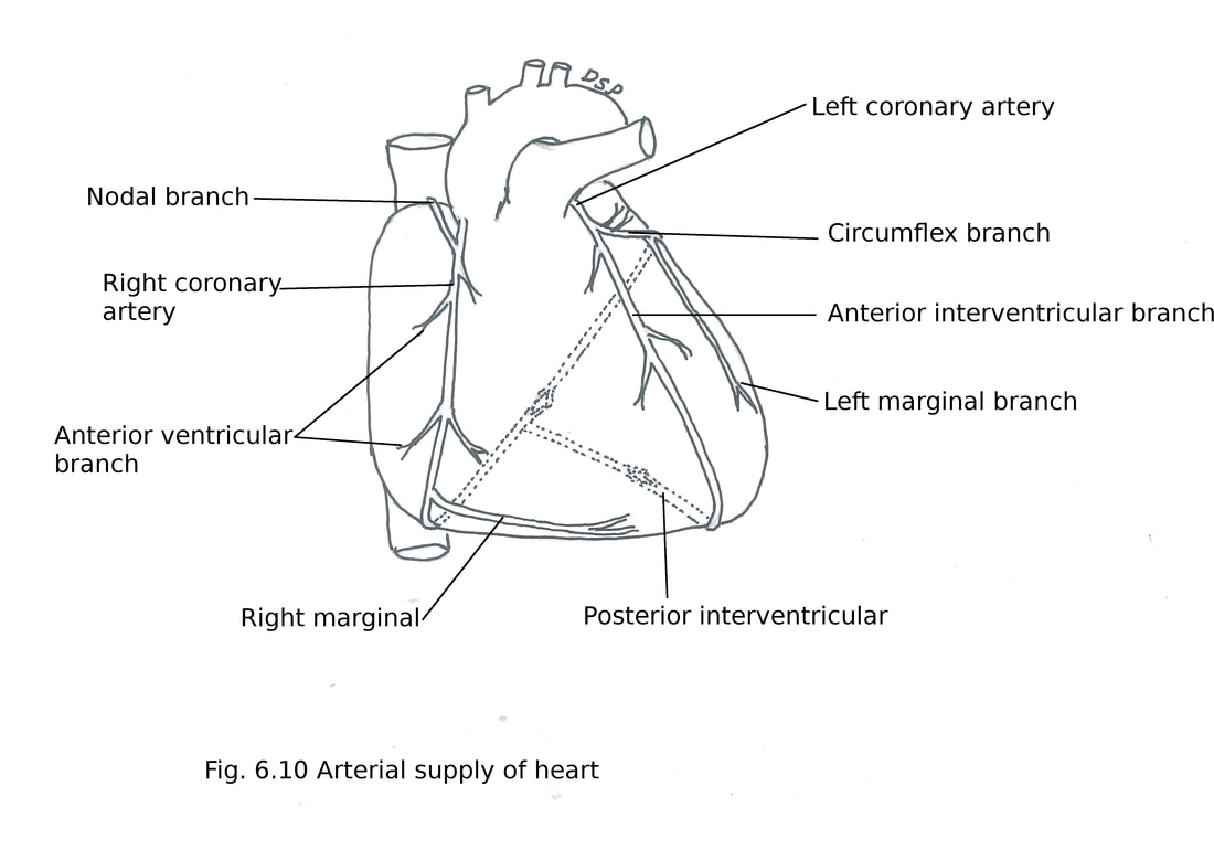
Arterial supply of Heart
The heart is supplied by two coronary arteries right and left. These two coronary arteries are branches of ascending aorta.
Right Coronary Artery :
It is dominant artery in about 60% people (posterior interventricular branch) supplying posterior part of ventricular septum part of posterolateral wall of left ventricle. It is a branch from anterior aortic sinus of ascending aorta.
It first goes forwards and to the right between right auricle and pulmonary trunk. Then runs downwards along atrioventricular groove up to inferior borders of the heart. Then winds round the inferior border along posterior part of atrioventricular groove reaches diaphragmatic surface of the heart up to junction of interatrial and interventricular grooves. Here it anastomoses with circumflex branch of left coronary artery.
Branches :
(1) Right marginal branch along inferior border of heart.
(2) Posterior interventricular branch near crux of heart lies in posterior interventricular groove. Near apex of heart anastomoses with anterior interventricular branch of left coronary artery. It gives septal branches to supply posteroinferior one third part of ventricular septum.
(3) Nodal branch supply sinuatrial node in about 60 to 65 % persons.
(4) Anterior atrial branches or rami arranged in anterior, lateral and posterior groups to supply right atrium.
(5) Infundibular branch supply infundibulum of right ventricle.
(6) Anterior ventricular branches arise from trunk of right coronary artery three to four in number supply sternocostal part of right ventricle.
(7) Posterior ventricular branches from trunk of right coronary artery lies in posterior part of atrioventricular groove. It supply diaphragmatic surface of right ventricle.
Areas of supplied by right coronary artery : Right atrium, part of left atrium, maximum part of right ventricle (excluding area adjoining anterior interventricular groove), part of the left ventricle diaphragmatic surface, posteroinferior one third part of the interventricular septum, conducting system of the heart (except a part of the left branch of the AV bundle).
Left Coronary Artery
It is a larger and wider branch than right coronary artery. It shows origin from left posterior aortic sinus of ascending aorta.
It goes forwards and left between left auricle and pulmonary trunk. It divides into two branches here anterior interventricular branch and circumflex branch.
Branches :
(1) Anterior interventricular branch is continuation of left coronary artery. It goes downwards in anterior interventricular groove on sternocostal surface of heart towards apex. After winding round reaches inferior surface into posterior interventricular groove and anastomoses with posterior interventricular branch of right coronary artery. It gives small branches anterior ventricular rami to supply both right and left ventricles. Anterior septal branches to supply anterosuperior two third part of ventricular septum.
(2) Circumflex branch goes along left part of atrioventricular groove. After winding round left border goes to posterior part of atrioventricular groove. Then it anastomoses with right coronary artery. It gives nodal branch to supply sinuatrial node in about 30 to 35 % persons. Left marginal branch runs along left border of heart towards apex supplying adjacent part of left ventricle to apex. Posterior interventricular branch is continuation of circumflex artery.
(3) Branches to left ventricle (diaphragmatic surface)
(4) Branches to left atrium
Areas of supplied by left coronary artery : Left atrium, ventricles in which maximum part of left ventricle (excluding area adjoining posterior interventricular groove), part of the right ventricle adjoining anterior interventricular groove, anterosuperior two third part of the interventricular septum, conducting system of the heart (a part of the left branch of the AV bundle).
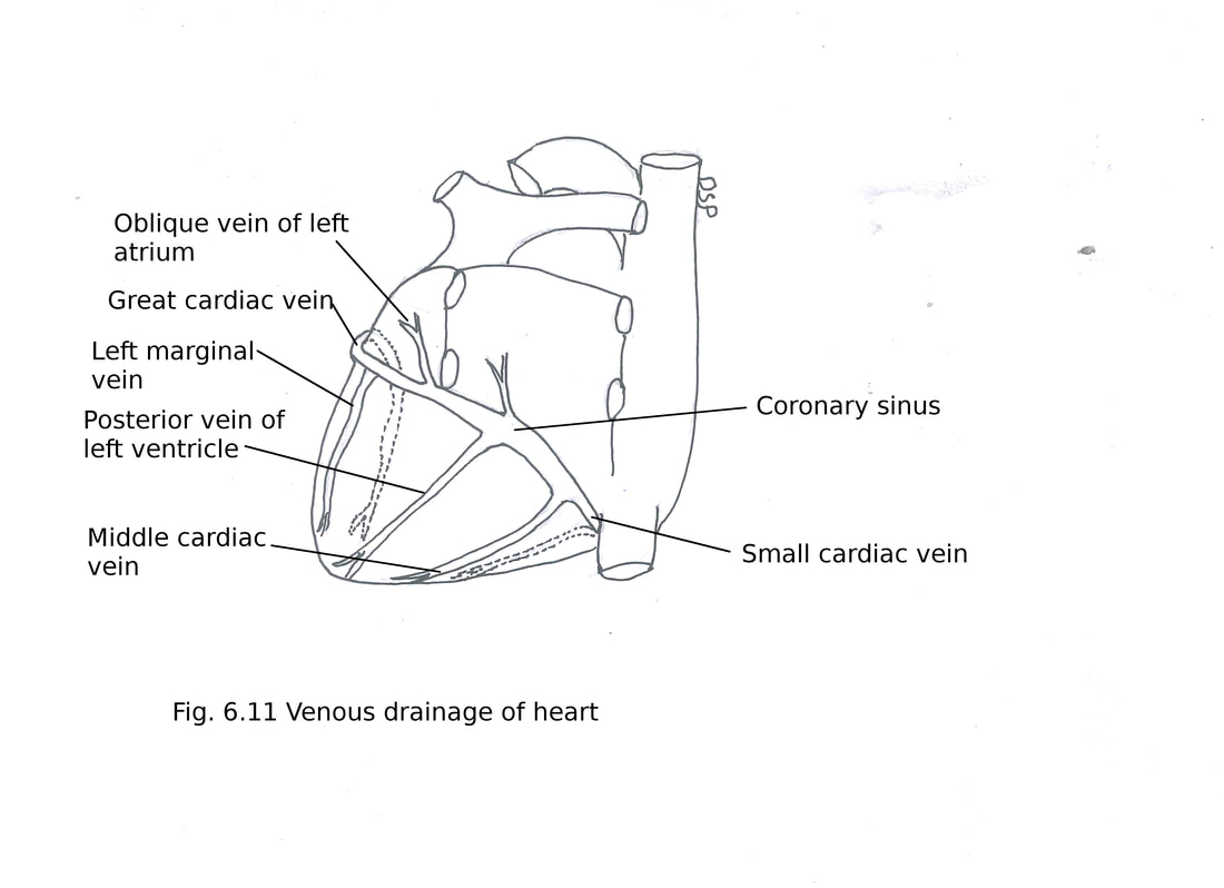
Venous drainage of Heart
Coronary sinus drains maximum venous blood of heart through its tributaries except anterior region of right ventricle and small part of right, left atrium and left ventricle. Whereas remaining small part drained by the venae cordis minimi (Thebesian veins) and anterior cardiac veins( anterior region of right ventricle).
Coronary sinus : This is the major vein of the heart. It is about 3 cm long situated in posterior atrioventricular groove.
Course : It starts from left part of the posterior atrioventricular groove. Then goes downwards and right in posterior atrioventricular groove. Finally it opens in right atrium between opening of inferior vena cava and right atrioventricular orifice. There is a presence of a semilunar valve near its opening in right atrial chamber.
Tributaries :
1. Great cardiac vein : It starts from apex of the heart goes upwards in anterior interventricular groove. Then turn around the left border. Finally opens in coronary sinus. It receives blood from left atrium, both ventricles.
2. Middle cardiac vein : It starts from the apex in posterior interventricular groove. After passing in relation with posterior interventricular groove opens in right side of coronary sinus.
3. Small cardiac vein : It lies in right posterior coronary sulcus between the right atrium and right
ventricle. Finally drains into coronary sinus near its right end. Right marginal vein joins with small cardiac vein for its drainage. It drains blood from posterior part of right atrium and right ventricle.
4. Posterior vein of the left ventricle : It is present on diaphragmatic surface of left ventricle. Drains into middle part of coronary sinus.
5. Oblique vein of the left atrium : It is a vein present on posterior surface of the left atrium. It drains into left end of coronary sinus.
Remaining veins which do not join with coronary sinus are as follows
6. Anterior cardiac veins : The are 3 to 4 small veins present on anterior surface of right ventricle. These small veins opens into the right.
7. Venae cordis minimi (Thebesian veins or small cardiac veins) : These are small veins present on all four chambers of heart. These open directly in to respective chambers of the heart through
foramina minimarum. These are more on right side of heart but less in number on left side.
8. Right marginal vein : It is a small vein which runs alongwith marginal branch of right coronary artery. It drains into small cardiac vein or right atrium directly.
Lymphatic drainage of the heart
Lymphatics vessels of heart forms subendocardial, myocardial and subepicardial network of plexuses. These vessels runs alongwith coronary arteries and form two trunks right and left. Right trunk goes to inferior brachiocephalic nodes and the left goes to inferior tracheobronchial lymph nodes.
Nerve supply of the heart
Heart is supplied by parasympathetic and sympathetic nerves. Both of these fibres are derived from cardiac plexus. Parasympathetic nerves are vagus. It decreases heart rate and constricts coronary arteries. Sympathetic nerves are derived from the upper 3 to 5 thoracic segments
of the spinal cord. It increases heart rate and dilates coronary arteries.
Parasympathetic and sympathetic nerves fibres form cardiac plexuses.
The superficial cardiac plexus :
It lies below arch of the aorta and anterior to right pulmonary artery. It is formed by the left superior cervical sympathetic ganglion its cardiac branch and lower two cervical cardiac branches of the left vagus nerve. It gives branches to the deep cardiac plexus, right coronary plexus and to left anterior pulmonary plexus.
The deep cardiac plexus:
It lies in front of bifurcation of the trachea, posterior to arch of the aorta and just above division of pulmonary trunk. It is formed by cardiac branches derived from superior, middle and inferior cervical ganglia and upper thoracic ganglia of the sympathetic chain. It also receive branches from vagus and recurrent laryngeal nerves. Only branches which go towards superficial plexus don't join here. On respective right and left side it gives branches to the corresponding coronary
and pulmonary plexuses. It gives small branches to right and left atria.
Applied anatomy :
1. Tachycardia : Increase in heart rate.
2. Bradycardia : Decrease in heart rate.
3. Fibrillation : Irregular, rapid twitching movement of muscles of heart. It may be atrial fibrillation or ventricular fibrillation.
4. Angina pectoris : This is pain in retrosternal region which radiate to inner side of left arm.
5. Myocardial infarction : Death of myocardial tissues due to sudden sudden and complete obstruction of any branch of coronary artery.
6. IHD Ischaemic heart disease : Tthis may due slow obstruction of any branch of coronary artery. This is due to formation of plaque in wall of coronary artery.
7. Congestive cardiac failure (CCF) : Failure of chamber of heart to pump blood efficiently. Causing increased venous return, edema feet, breathlessness. Right sides or left sided failure may be there.
