GLUTEAL REGION
Extent
Superiorly : Iliac crest
Inferiorly : Gluteal fold
Anteriorly : Anterior superior iliac spine to anterior part of greater trochanter
Poteriorly : Sacral spines
Superficial fascia
It is thick containing fat. Fat is more abundant in adult female giving rounded contour to gluteal region.
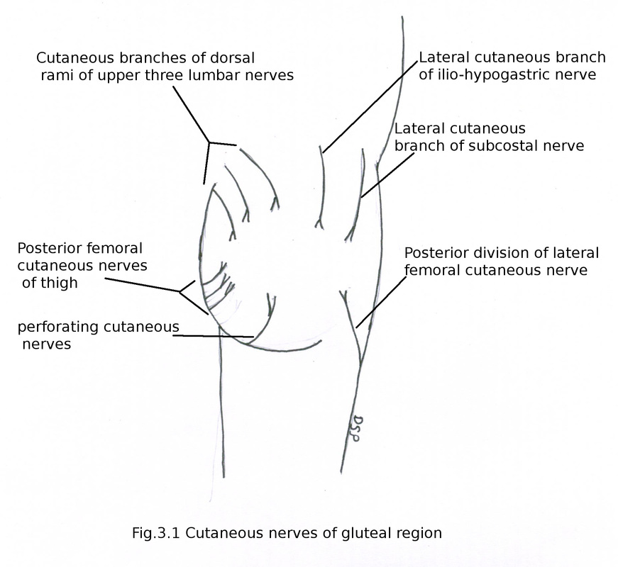
Cutaneous nerves in superficial fascia present.
These are as follows :
1) Anterior and upper quadrant : Lateral cutaneous branch of subcostal nerve (T12), lateral cutaneous branch of ilio-hypogastric nerve.
2) Anterior and lower quadrant : Posterior division of lateral femoral cutaneous nerve (L2,L3).
3) Posterior and upper quadrant : Cutaneous branches of dorsal rami of upper three lumbar nerves (L1,L2,L3).
4) Posterior and lower quadrant : Posterior femoral cutaneous nerves of thigh (S1,S2,S3), perforating cutaneous nerves (S2,S3).
Extent
Superiorly : Iliac crest
Inferiorly : Gluteal fold
Anteriorly : Anterior superior iliac spine to anterior part of greater trochanter
Poteriorly : Sacral spines
Superficial fascia
It is thick containing fat. Fat is more abundant in adult female giving rounded contour to gluteal region.
Cutaneous nerves in superficial fascia present.
These are as follows :
1) Anterior and upper quadrant : Lateral cutaneous branch of subcostal nerve (T12), lateral cutaneous branch of ilio-hypogastric nerve.
2) Anterior and lower quadrant : Posterior division of lateral femoral cutaneous nerve (L2,L3).
3) Posterior and upper quadrant : Cutaneous branches of dorsal rami of upper three lumbar nerves (L1,L2,L3).
4) Posterior and lower quadrant : Posterior femoral cutaneous nerves of thigh (S1,S2,S3), perforating cutaneous nerves (S2,S3).
Deep fascia
It shows attachment on iliac crest above and behind on sacrum. It splits to enclose tensor fascia latae and gluteus maximus muscles. It forms gluteal aponeurosis a thick sheet covering gluteus medius. Layer which covers gluteus maximus are connected by interconnecting fibrous septa. Laterally deep fascia continue as ilio-tibial tract.
Muscles :
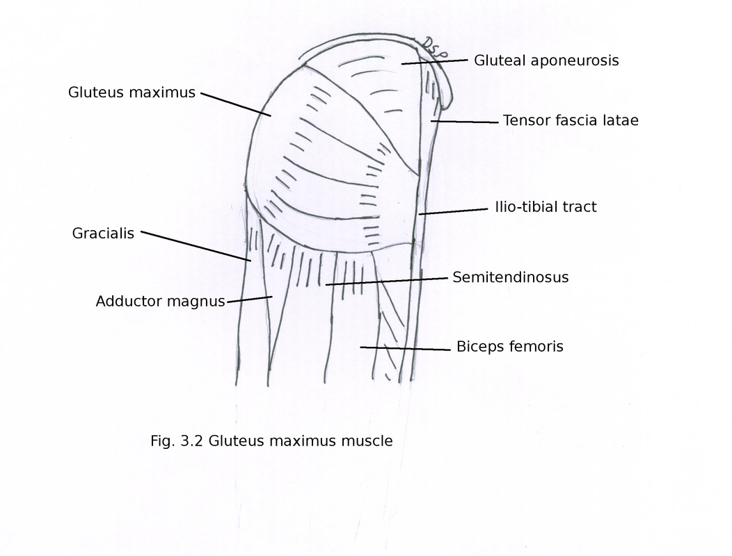
1) Gluteus maximus
It is big and superficial muscle of gluteal region.
Origin : It shows origin from gluteal surface of ilium behind posterior gluteal line, dorsal segment of iliac crest from outer part, from fascia covering erector spinae, from dorsal surface of lower part sacrum, from side of coccyx and from sacrotuberous ligament.
Insertion : It goes laterally and shows insertion of its one fourth part on gluteal tuberosity and three fourth fibres on ilio-tibial tract of fascia latae.
Nerve supply : It receives nerve supply from inferior gluteal (L5,S1,S2) nerve from its deep part.
Action : Extension of hip joint during extreme position like extension of flexed hip in climbing running but inactive during standing, help in elevation of trunk from forward flexed position , lateral rotation of hip, upper fibres help in powerful abduction of hip, lateral rotation of hip joint.
It maintain extended position of knee joint through ilio-tibial tract.
It shows attachment on iliac crest above and behind on sacrum. It splits to enclose tensor fascia latae and gluteus maximus muscles. It forms gluteal aponeurosis a thick sheet covering gluteus medius. Layer which covers gluteus maximus are connected by interconnecting fibrous septa. Laterally deep fascia continue as ilio-tibial tract.
Muscles :
1) Gluteus maximus
It is big and superficial muscle of gluteal region.
Origin : It shows origin from gluteal surface of ilium behind posterior gluteal line, dorsal segment of iliac crest from outer part, from fascia covering erector spinae, from dorsal surface of lower part sacrum, from side of coccyx and from sacrotuberous ligament.
Insertion : It goes laterally and shows insertion of its one fourth part on gluteal tuberosity and three fourth fibres on ilio-tibial tract of fascia latae.
Nerve supply : It receives nerve supply from inferior gluteal (L5,S1,S2) nerve from its deep part.
Action : Extension of hip joint during extreme position like extension of flexed hip in climbing running but inactive during standing, help in elevation of trunk from forward flexed position , lateral rotation of hip, upper fibres help in powerful abduction of hip, lateral rotation of hip joint.
It maintain extended position of knee joint through ilio-tibial tract.
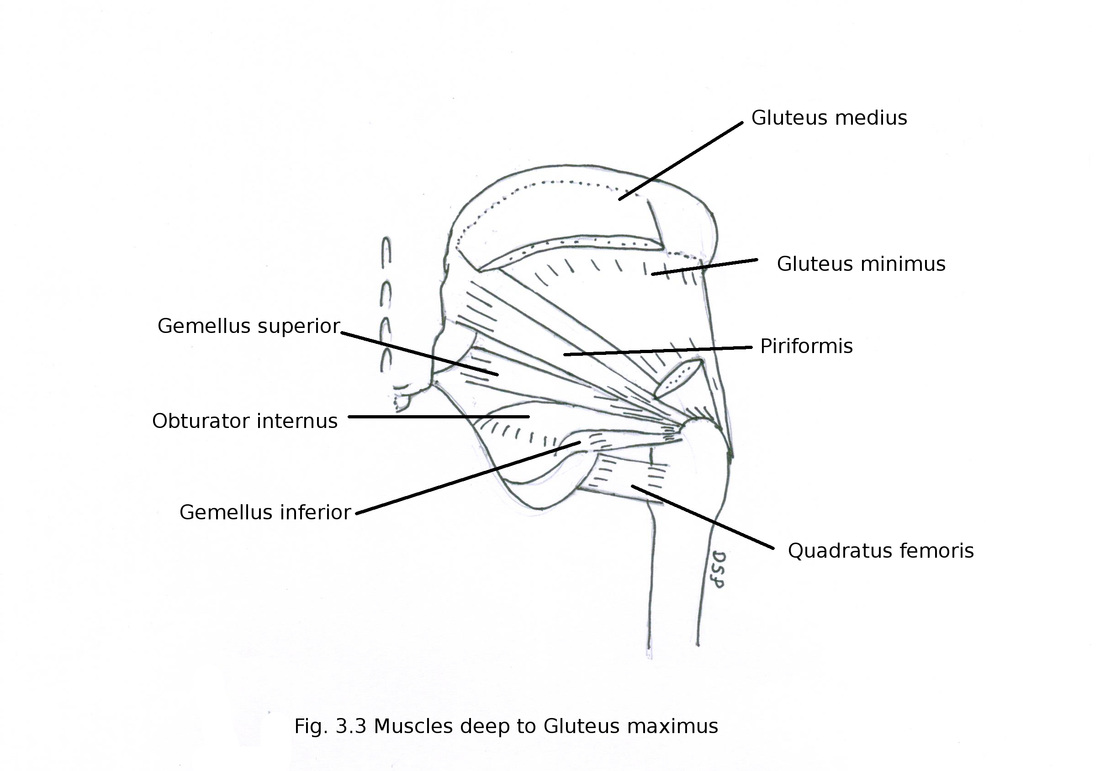
2) Gluteus medius
It is partly deep to gluteus maximus.
Origin : It shows origin from gluteal surface of ilium between posterior gluteal line posteriorly, anterior gluteal line anteriorly and iliac crest superiorly. Also from fascia covering it.
Insertion : It goes laterally, downward and anteriorly shows insertion on outer surface of greater trochanter on a line which goes downward and forward.
Nerve supply : It receives nerve supply from superior gluteal (L4,L5,S1) nerve.
Action : Abduction of hip joint. Medial rotation by anterior fibres. It acts from below alongwith gluteus minimus prevent sagging down of opposite unsupported side pelvis during walking.
3) Gluteus minimus
It lies deep to gluteus medius.
Origin : It shows origin from gluteal surface of ilium between anterior gluteal line and inferior gluteal line, along a line in relation to greater sciatic notch.
Insertion : It shows insertion by a tendon on outer surface of greater trochanter on anterior aspect. Nerve supply : It receives nerve supply from superior gluteal (L4,L5,S1) nerve.
Action : Abduction of hip joint. Medial rotation by anterior fibres. It acts from below alongwith gluteus minimus prevent sagging down of opposite unsupported side pelvis during walking.
Applied anatomy : Trendelenberg's sign in paralysis of gluteus medius and minimus pelvis goes down on unsupported side when person standing on affected limb.
4) Tensor fascia latae
Origin : It shows origin from outer lip of iliac crest about 5 cm behind anterior superior iliac spine to tubercle of iliac crest.
Insertion : It shows insertion on ilio-tibial tract of fascia latae.
Nerve supply : It receives nerve supply from superior gluteal (L4,L5,S1) nerve.
Action : Abduction of hip joint. It help in maintenance of extension of knee joint.
5) Piriformis
Origin : It shows origin from anterior surface of sacrum area between anterior sacral foramina, from gluteal surface of ilium adjacent to posterior inferior iliac spine, capsule of sacroiliac joint and sacrotuberous ligament.
Insertion : It goes laterally through greater sciatic foramen and shows insertion by a tendon on upper margin of greater trochanter.
Nerve supply : It receives nerve supply from L5, S1, S2 nerve.
Action : Abduction of hip joint while it is in flexed position. It act as lateral rotator of hip joint.
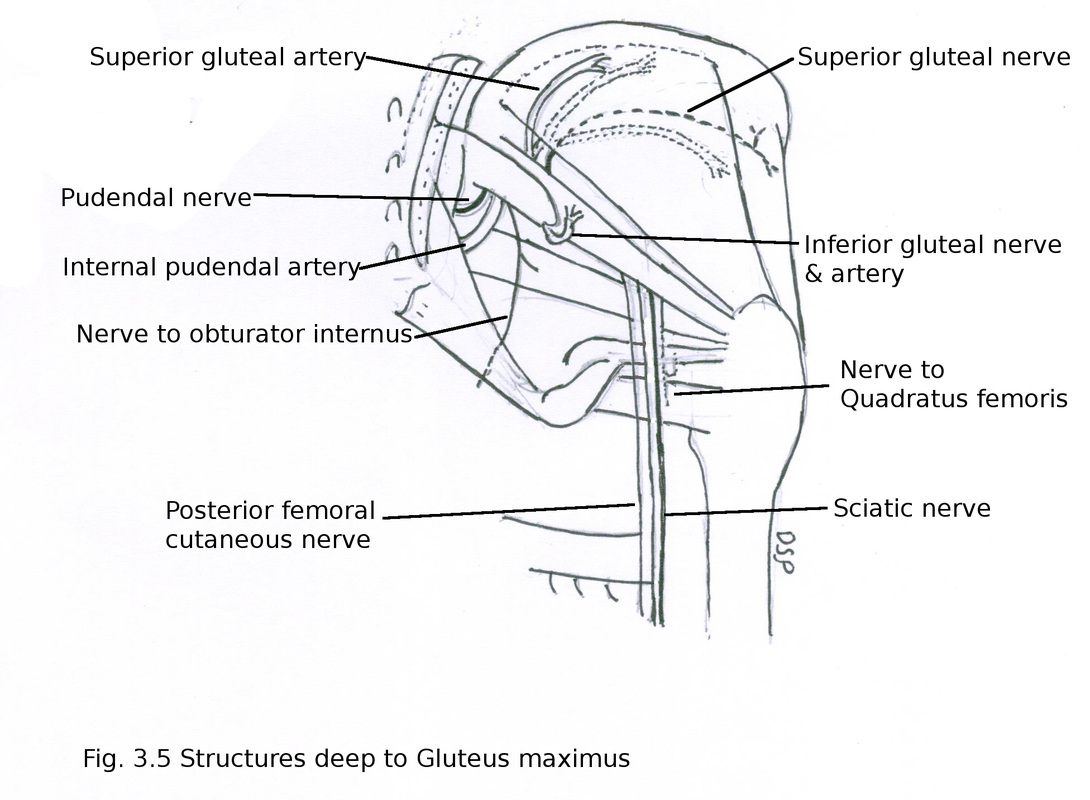
Structures lying in relation to upper margin of piriformis :
superior gluteal vessels and nerve
Structures lying in relation to lower margin of piriformis (lateral to medial side) :
a) Sciatic nerve and nerve to quadratus femoris deep to it
b) Posterior femoral cutaneous nerve
c) Inferior gluteal vessels and nerve
d) Nerve to obturator internus
e) Internal pudendal vessels
f) Pudendal nerve
6) Obturator internus
Origin : It shows origin on inner surface of lesser pelvis around obturator foramen from ischio-pubic ramus, body of ischium, area between below pelvic brim and greater sciatic foramen. It also shows origin from inner surface of obturator membrane.
Insertion : Its fibres joins and turn around between ischial spine and tuberosity of ischium goes out of pelvis through lesser sciatic foramen two gemelli joins with it and shows insertion on medial surface of greater trochanter anterior to trochanteric fossa.
Nerve supply : It receives nerve supply from nerve to obturator internus L5, S1.
Action : It act as lateral rotator of hip in extended position of thigh and abductor of hip joint in flexed position of thigh.
7) Gemellus superior
Origin : It shows origin from dorsal surface of ischial spine.
Insertion : Its fibres joins with upper margin of obturator internus and shows insertion on medial surface of greater trochanter anterior to trochanteric fossa.
Nerve supply : It receives nerve supply from nerve to obturator internus L5, S1.
Action : It act as lateral rotator of hip in extended position of thigh and abductor of hip joint in flexed position of thigh.
8) Gemellus inferior
Origin : It shows origin from ischial tuberosity from its upper part near lower margin of lesser sciatic notch.
Insertion : Its fibres joins with lower margin of obturator internus and shows insertion on medial surface of greater trochanter anterior to trochanteric fossa.
Nerve supply : It receives nerve supply from nerve to quadratus femoris L4,L5,S1.
Action : It act as lateral rotator of hip in extended position of thigh and abductor of hip joint in flexed position of thigh.
9) Quadratus femoris
It lies below gemellus inferior.
Origin : It shows origin from outer surface of upper part of ischial tuberosity.
Insertion : Its fibres goes laterally on quadrate tubercle in middle part of inter-trochanteric crest.
Nerve supply : It receives nerve supply from nerve to quadratus femoris L4,L5,S1.
Action : It act as lateral rotator of hip.
It is partly deep to gluteus maximus.
Origin : It shows origin from gluteal surface of ilium between posterior gluteal line posteriorly, anterior gluteal line anteriorly and iliac crest superiorly. Also from fascia covering it.
Insertion : It goes laterally, downward and anteriorly shows insertion on outer surface of greater trochanter on a line which goes downward and forward.
Nerve supply : It receives nerve supply from superior gluteal (L4,L5,S1) nerve.
Action : Abduction of hip joint. Medial rotation by anterior fibres. It acts from below alongwith gluteus minimus prevent sagging down of opposite unsupported side pelvis during walking.
3) Gluteus minimus
It lies deep to gluteus medius.
Origin : It shows origin from gluteal surface of ilium between anterior gluteal line and inferior gluteal line, along a line in relation to greater sciatic notch.
Insertion : It shows insertion by a tendon on outer surface of greater trochanter on anterior aspect. Nerve supply : It receives nerve supply from superior gluteal (L4,L5,S1) nerve.
Action : Abduction of hip joint. Medial rotation by anterior fibres. It acts from below alongwith gluteus minimus prevent sagging down of opposite unsupported side pelvis during walking.
Applied anatomy : Trendelenberg's sign in paralysis of gluteus medius and minimus pelvis goes down on unsupported side when person standing on affected limb.
4) Tensor fascia latae
Origin : It shows origin from outer lip of iliac crest about 5 cm behind anterior superior iliac spine to tubercle of iliac crest.
Insertion : It shows insertion on ilio-tibial tract of fascia latae.
Nerve supply : It receives nerve supply from superior gluteal (L4,L5,S1) nerve.
Action : Abduction of hip joint. It help in maintenance of extension of knee joint.
5) Piriformis
Origin : It shows origin from anterior surface of sacrum area between anterior sacral foramina, from gluteal surface of ilium adjacent to posterior inferior iliac spine, capsule of sacroiliac joint and sacrotuberous ligament.
Insertion : It goes laterally through greater sciatic foramen and shows insertion by a tendon on upper margin of greater trochanter.
Nerve supply : It receives nerve supply from L5, S1, S2 nerve.
Action : Abduction of hip joint while it is in flexed position. It act as lateral rotator of hip joint.
Structures lying in relation to upper margin of piriformis :
superior gluteal vessels and nerve
Structures lying in relation to lower margin of piriformis (lateral to medial side) :
a) Sciatic nerve and nerve to quadratus femoris deep to it
b) Posterior femoral cutaneous nerve
c) Inferior gluteal vessels and nerve
d) Nerve to obturator internus
e) Internal pudendal vessels
f) Pudendal nerve
6) Obturator internus
Origin : It shows origin on inner surface of lesser pelvis around obturator foramen from ischio-pubic ramus, body of ischium, area between below pelvic brim and greater sciatic foramen. It also shows origin from inner surface of obturator membrane.
Insertion : Its fibres joins and turn around between ischial spine and tuberosity of ischium goes out of pelvis through lesser sciatic foramen two gemelli joins with it and shows insertion on medial surface of greater trochanter anterior to trochanteric fossa.
Nerve supply : It receives nerve supply from nerve to obturator internus L5, S1.
Action : It act as lateral rotator of hip in extended position of thigh and abductor of hip joint in flexed position of thigh.
7) Gemellus superior
Origin : It shows origin from dorsal surface of ischial spine.
Insertion : Its fibres joins with upper margin of obturator internus and shows insertion on medial surface of greater trochanter anterior to trochanteric fossa.
Nerve supply : It receives nerve supply from nerve to obturator internus L5, S1.
Action : It act as lateral rotator of hip in extended position of thigh and abductor of hip joint in flexed position of thigh.
8) Gemellus inferior
Origin : It shows origin from ischial tuberosity from its upper part near lower margin of lesser sciatic notch.
Insertion : Its fibres joins with lower margin of obturator internus and shows insertion on medial surface of greater trochanter anterior to trochanteric fossa.
Nerve supply : It receives nerve supply from nerve to quadratus femoris L4,L5,S1.
Action : It act as lateral rotator of hip in extended position of thigh and abductor of hip joint in flexed position of thigh.
9) Quadratus femoris
It lies below gemellus inferior.
Origin : It shows origin from outer surface of upper part of ischial tuberosity.
Insertion : Its fibres goes laterally on quadrate tubercle in middle part of inter-trochanteric crest.
Nerve supply : It receives nerve supply from nerve to quadratus femoris L4,L5,S1.
Action : It act as lateral rotator of hip.
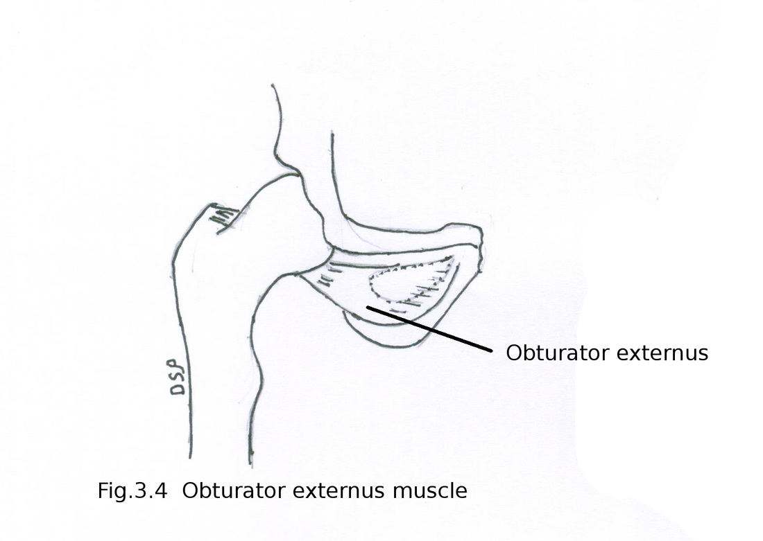
10) Obturator externus
Origin : It shows origin from on anterior surface of obturator membrane alongwith medial margin of obturator foramen and part of pubis, ischial ramus around it except upper part near obturator notch.
Insertion : It forms a tendon which goes backward, laterally and upwards on the back of neck of femur. It shows insertion on trochanteric fossa of femur.
Nerve supply : It receives nerve supply from posterior division of obturator nerve L3, L4.
Action : It is a postural muscle. Lateral rotator of hip joint.
Nerves :
1. Superior gluteal nerve
Formed by dorsal branches of ventral rami of L4,L5,S1. It passes through greater sciatic foramen above piriformis. It divides in to superior and inferior branches to supply gluteus medius and gluteus minimus. Inferior branch also supply tensor fascia latae.
2. Inferior gluteal nerve
Formed by dorsal branches of ventral rami of L5,S1,S2. It passes through greater sciatic foramen below piriformis. It supply gluteus maximus from its deep aspect.
3. Nerve to quadratus femoris and gemellus inferior
Formed by ventral branches of ventral rami of L4,L5,S1. It passes through greater sciatic foramen below piriformis deep to sciatic nerve, gemelli and obturator internus tendon. It supply quadratus femoris, gemellus inferior and give a branch to hip joint.
4. Posterior femoral cutaneous nerve
Formed by dorsal branches of S1,S2 and ventral branches of S2,S3. It passes through greater sciatic foramen below piriformis medial to sciatic nerve. It goes down to back of thigh deep to fascia latae. In relation with roof of popliteal fossa accompanies small saphenous vein and communicate with sural nerve.
Branches 1) Gluteal : Supply skin of lower and lateral part of gluteal region. 2) Perineal : Supply superomedial skin of thigh and posterior part of scrotum or labium majus. 3) Perforating : Supply skin of back of thigh, popliteal fossa and upper part of back of leg.
5. Nerve to obturator internus and gemellus superior
Formed by ventral branches of ventral rami of L5,S1,S2. It passes through greater sciatic foramen below piriformis on surface of ischial spine. Then passes into lesser sciatic foramen. It supply obturator internus also gemellus superior.
6. Pudendal nerve
Formed by ventral branches of ventral rami of S2,S3,S4. It passes through greater sciatic foramen below piriformis medial to ischial spine. Then passes into lesser sciatic foramen. It supply perineal region after passing through pudendal canal. It gives branches inferior rectal nerve, perineal nerve, dorsal nerve of penis or clitoris.
7. Sciatic nerve
It is thick nerve about 2 cm thick. It shows two components tibial and common peroneal. Tibial component formed by ventral branches of ventral rami of L4,L5,S1,S2,S3 and common peroneal component formed by dorsal branches of ventral rami of L4,L5,S1,S2 . It passes through greater sciatic foramen below piriformis a mid point between ischial tuberosity and greater trochanter. Then passes down over the surface of dorsal surface of body of ischium, obturator internus with gemellus superior and inferior, quadratus femoris, adductor magnus. Here it is accompanied by posterior femoral cutaneous nerve and inferior gluteal artery. Near upper angle of popliteal fossa it gives out tibial and common peroneal nerves.
Branches : These are muscular branches to long head of biceps femoris, ischial part of adductor magnus, semitendinosus, semimembranosus and a branch to hip joint.
Applied anatomy : Injury to sciatic nerve by fracture with dislocation of femur, penetrating wound at back of thigh is common. When injury occurs muscles of leg below knee joint get paralyzed, cutaneous loss of all sensations sparing area supplied by saphenous nerve causing foot drop. Partial injury to nerve may be caused by continue external pressure on nerve by hard surface, injections in gluteus maximus etc.
8. Perforating cutaneous nerve of thigh
Formed by ventral rami of S2,S3. After piercing sacrotuberous passes below lower margin of gluteus maximus to supply inferior and medial aspect of gluteal region.
Blood vessels :
1. Superior gluteal artery
It is a branch of internal iliac artery from its posterior division. Alongwith superior gluteal nerve goes to gluteal region through greater sciatic foramen in relation to upper margin of piriformis. Its branches are superficial and deep. Superficial branch lies in between gluteus maximus and gluteus medius muscles. Deep branch lies in between gluteus medius and gluteus minimus muscles divides into upper and lower branches. Spinous and trochanteric anastomosis formed by its branches.
Spinous anastomosis
Formed by superficial circumflex iliac, deep circumflex iliac artery, ascending branch of lateral circumflex femoral artery, iliac branch of ilio-lumbar artery and upper branch of deep branch of superior gluteal artery. This anastomosis is in relation to anterior superior iliac spine.
Trochanteric anastomosis
Formed by lower branch of deep branch of superior gluteal artery, ascending branch of lateral circumflex femoral artery, ascending branch medial circumflex femoral artery, branch of inferior gluteal artery. This anastomosis is in the trochanteric fossa.
2. Inferior gluteal artery
It is a branch of internal iliac artery from its anterior division. Alongwith inferior gluteal nerve goes to gluteal region in relation to lower margin of piriformis. Its branches are anastomotic, muscular, arteria nervi ischiadici. Anastomotic branches forms cruciate and trochanteric anastomosis, muscular branches supply muscles of region and arteria nervi ischiadici supply sciatic nerve.
Cruciate anastomosis
Formed by transverse branch of medial circumflex femoral artery,descending branch of inferior gluteal artery, ascending branch of 1st perforating artery and transverse branch of lateral circumflex femoral artery. It is formed near greater trochanter of femur.
3. Internal Pudendal artery
It is a branch of internal iliac artery from its anterior division below origin of obturator artery. Goes to gluteal region in relation to lower margin of piriformis. Alongwith venae comitantes comes in relation with dorsal surface of ischial spine. Here lies nerve to obturator internus laterally and pudendal nerve medially. Then it passes through lesser sciatic foramen and goes to pudendal canal. It gives muscular branches to supply muscles of perineum and gluteal region.
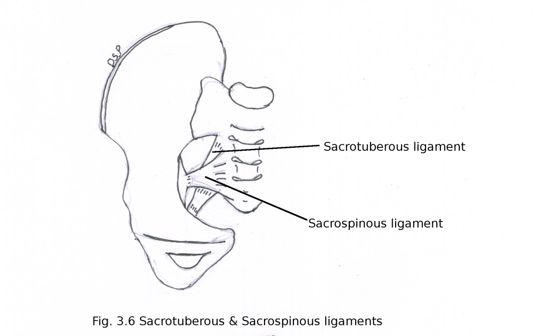
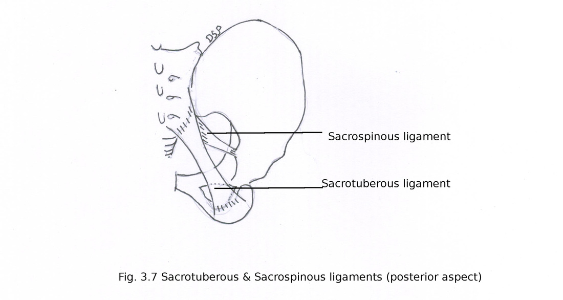
Ligaments :
Sacrotuberous ligament
It is fibrous band shows attachment medially on posterior superior iliac spine, posterior inferior iliac spine, transverse tubercle on lower part, lateral border of sacrum lower part and upper part of coccyx. Laterally shows attachment on medial margin of ischial tuberosity.
Part of gluteus maximus shows attachment on posterior surface.
Sacrospinous ligament
It is triangular ligament shows attachment on lateral margin of sacrum and coccyx. Laterally shows attachment on ischial spine.
Sacrotuberous and sacrospinous ligaments forms greater and lesser sciatic foramina.