FOREARM
Forearm has 2 compartments
1. Anterior 2. Posterior
1. Anterior or flexor compartment
Contents : Muscles (superficial group and deep group), vessels (radial and ulnar artery and their branches), nerves (median nerve, ulnar nerve and radial nerve).
Muscles superficial group and deep group.
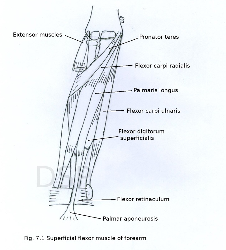
Superficial muscles are lateral to medial 5
1. Pronator teres
Origin: Humeral head lies superficial and shows origin on medial epicondyle of humerus and medial supracondylar ridge. Ulnar head lies deep and shows origin from medial margin of coronoid process of ulna.
Insertion: Both head joins with each other goes downwards and laterally and shows insertion on middle part of lateral surface of radius.
Nerve supply: from media nerve.
Action : Pronation of forearm and flexion at elbow joint.
2. Flexor carpi radialis
Origin: It shows origin on medial epicondyle of humerus.
Insertion : It has two tendon shows insertion on base of second and third metacarpal bones.
Nerve supply: Median nerve.
Action: Flexion at wrist joint, abduction at wrist joint.
3. Palmaris longus
Origin : It shows origin from medial epicondyle of humerus.
Insertion: This muscle continue as palmar aponeurosis in hand near apex of palmar aponeurosis.
Nerve supply : Median nerve.
Action : Flexion at wrist joint.
4. Flexor carpi ulnaris
Origin : Humeral head shows origin from medial epicondyle of humerus and ulnar head shows origin from medial margin of olecranon process and upper part of posterior border of ulna.
Insertion : Tendon of muscle shows insertion on pisiform bone and with the help of pisohamate ligament and pisometacarpal ligament shows insertion on hook of hamate and base of fifth metacarpal bone respectively.
Action : Flexion at wrist joint and adduction at wrist joint.
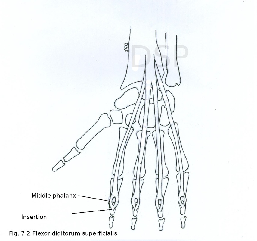
5. Flexor digitorum superficialis
Origin : Humeroulnar head shows origin from medial epicondyle of humerus and anterior part of ulnar collateral ligament and also from medial margin coronoid process of ulna, radial head shows origin from anterior border of radius which is oblique.
Insertion : This muscles divided into 4 tendons for medial 4 fingers. These tendons split into 2 parts near proximal phalanx and finally shows insertion on shaft of of middle phalanx of medial 4 digits.
Nerve supply : median nerve.
Action : Muscle produces flexion at proximal interphalangeal joint of medial four fingures.
Forearm has 2 compartments
1. Anterior 2. Posterior
1. Anterior or flexor compartment
Contents : Muscles (superficial group and deep group), vessels (radial and ulnar artery and their branches), nerves (median nerve, ulnar nerve and radial nerve).
Muscles superficial group and deep group.
Superficial muscles are lateral to medial 5
- Pronator teres
- Flexor carpi radialis
- Palmaris longus
- Flexor carpi ulnaris
- Flexor digitorum superficialis
- Flexor pollicis longus
- Flexor digitorum profundus
- Pronator quadratus
1. Pronator teres
Origin: Humeral head lies superficial and shows origin on medial epicondyle of humerus and medial supracondylar ridge. Ulnar head lies deep and shows origin from medial margin of coronoid process of ulna.
Insertion: Both head joins with each other goes downwards and laterally and shows insertion on middle part of lateral surface of radius.
Nerve supply: from media nerve.
Action : Pronation of forearm and flexion at elbow joint.
2. Flexor carpi radialis
Origin: It shows origin on medial epicondyle of humerus.
Insertion : It has two tendon shows insertion on base of second and third metacarpal bones.
Nerve supply: Median nerve.
Action: Flexion at wrist joint, abduction at wrist joint.
3. Palmaris longus
Origin : It shows origin from medial epicondyle of humerus.
Insertion: This muscle continue as palmar aponeurosis in hand near apex of palmar aponeurosis.
Nerve supply : Median nerve.
Action : Flexion at wrist joint.
4. Flexor carpi ulnaris
Origin : Humeral head shows origin from medial epicondyle of humerus and ulnar head shows origin from medial margin of olecranon process and upper part of posterior border of ulna.
Insertion : Tendon of muscle shows insertion on pisiform bone and with the help of pisohamate ligament and pisometacarpal ligament shows insertion on hook of hamate and base of fifth metacarpal bone respectively.
Action : Flexion at wrist joint and adduction at wrist joint.
5. Flexor digitorum superficialis
Origin : Humeroulnar head shows origin from medial epicondyle of humerus and anterior part of ulnar collateral ligament and also from medial margin coronoid process of ulna, radial head shows origin from anterior border of radius which is oblique.
Insertion : This muscles divided into 4 tendons for medial 4 fingers. These tendons split into 2 parts near proximal phalanx and finally shows insertion on shaft of of middle phalanx of medial 4 digits.
Nerve supply : median nerve.
Action : Muscle produces flexion at proximal interphalangeal joint of medial four fingures.
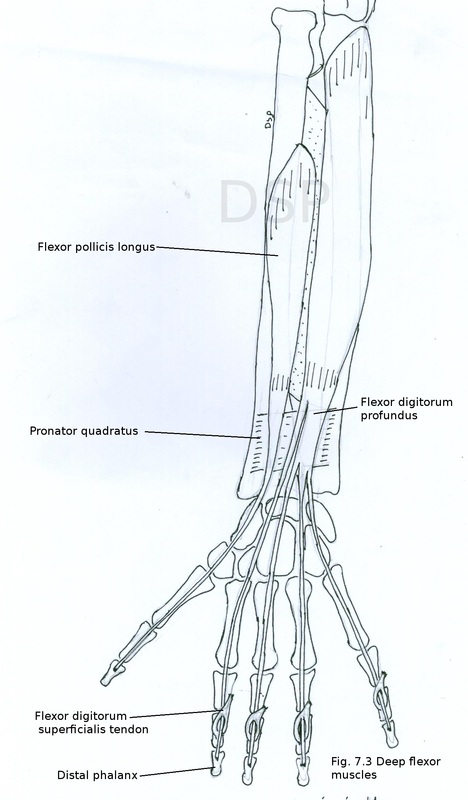
Deep flexor muscles
1. Flexor pollicis longus
Origin : It shows origin from anterior surface of shaft of radius below oblique part of anterior margin, anterior surface of intraosseous membrane and from medial margin of coronoid process of ulna.
Insertion : The muscle tendon passes deep to flexor retinaculum and shows insertion on palmar surface of distal phalanx of thumb.
Nerve supply : Anterior interosseous nerve branch of median nerve.
Action : It flexes distal interphalangeal joint of thumb.
2. Flexor digitotum profundus
Origin : It shows origin from upper 3/4th part of anterior and medial surface of shaft of ulna, medial surface of olecranon process and coronoid process, upper 3/4 part of anterior surface of interosseus membrane and from upper 3/4th part of posterior border of ulna by an aponeurosis and interosseus membrane in upper part. In lower part it shows four tendons for medial 4 fingers.
Insertion : Its tendons lies deep to flexor retinaculum near proximal phalanx tendons passes through tendon of flexor digitorum superficialis. Finally shows insertion on palmar surface of base of distal phalanx of medial 4 fingers.
Nerve supply : Medial part of muscle receives nerve supply from ulnar nerve and lateral part receives nerve supply from anterior interosseus nerve which is branch of median nerve C8, T1.
Action : Flexes distal phalanges when middle phalanges flexed by flexor digitorum superficialis. Flexes middle phalanx. It also help in flexion at metacarpophalangeal and intercarpal joints.
3. Pronator quadratus
Origin : It shows origin from oblique ridge on lower part of anterior surface of ulna. It shows two fibres superficial and deep. Superficial fibres goes downwards and laterally.
Insertion : Superficial fibers shows insertion on anterior surface of lower part of radius. Deep part shows insertion on triagular area above ulnar notch.
Nerve supply : From anterior interosseus nerve which is branch of median nerve.
Action : Superficial fibres help in pronation of forearm. Deep fibres help in binding of radius and ulna in lower part.
Vessels
Radial and ulnar arteries are vessels of 4 forearm. These are branches of brachial artery in cubital fossa.
Radial artery
It is a small branch of brachial artery. It originates in cubital fossa medial to tendon of biceps. In forearm it goes downwards with slight convexity laterally. In upper part of forearm covered by brachioradialis and in lower part covered by skin and fascia. This artery present over surface of biceps tendon, supinator muscle, pronator teres muscle, flexor digitorum superficialis muscle, flexor pollicis longus muscle, pronator quadratus muscle and anterior surface of lower end of radius from above downwards. At wrist joint it goes dorsally into anatomical snuff box after passing between two heads of first dorsal interosseous muscle. It enters the palm to join with deep branch of ulnar artery to form deep palmar arch.
Branches
Pulsations of radial artery can be felt in lower anterior part of radius.
Ulnar artery
It is large branch of brachial artery arising in cubital fossa.
Course : In upper part it runs downward and medially. Then it runs vertically downwards to wrist joint. During its course lies over the surface of brachialis and flexor digitorum profundus muscles. Anteriorly it is covered by pronator teres muscle, flexor carpi radialis muscle, palmaris longus muscle, flexor digitorum superficial muscles and flexor carpi ulnaris muscle from above downwards. Part of artery in relation to radius lower part covered by skin, fascia only. Medially it is accompanied by ulnar nerve.
Branches
1. Anterior and posterior ulnar recurrent artery
2. Common interosseous artery
3. Palmar and dorsal carpal branches
4. Muscular branches
1. Anterior and posterior ulnar recurrent artery
Both these arteries goes upwards in relation to medial epicondyle of humerus anterior and posterior to it. Anastomoses with inferior ulnar collateral artery anteriorly and with ulnar collateral branch and interosseous recurrent artery posteriorly respectively.
2. Common interosseous artery
It arises in cubital fossa. It goes downwards backwards up to upper margin of interosseous membrane. It divides into two branches anterior and posterior interosseous artery.
Anterior interosseous artery runs downwards in close contact with anterior surface of interosseous membrane along with anterior interosseous branch of median nerve. In lower part of interosseous membrane along upper margin of pronator teres it penetrates interosseous membrane to anastomose with posterior interosseous artery. It supply muscles, radius and ulna .
Posterior interosseous artery runs downwards in close contact with interosseous membrane along with deep branch of radial nerve. Here it lies in between superficial and deep group of extensor muscle in lower part. It gives a branch interosseous recurrent artery which anastomoses with middle collateral branch of profunda brachii artery posterior to lateral epicondyle.
3. Palmar and dorsal carpal branches
It helps in formation of anastomosis around wrist joint. Palmar carpal branch form palmar carpal arch and dorsal carpal branch form dorsal carpal arch.
4. Muscular branches
Muscular branches supply all muscles of forearm.
Nerves of the front of forearm
Nerves of front of forearm are 1. Median nerve 2. Ulnar nerve and 3. Radial nerve.
1. Median nerve
It is a nerve of the front of forearm. It lies medial to brachial artery in cubital fossa. It passes between two heads of pronator teres muscle and goes to front of forearm. Deep to deep head of pronator teres lies ulnar artery. Then it runs over surafce of flexor digitorum profundus deep to humeroulnar and radial heads of flexor digitorum superficialis. It passes between superficial and deep groups of flexor muscles. It goes downwards towards wrist on posterior surface of flexor digitorum superficialis. Just 5 cm above flexor retinaculum it becomes superficial after passing laterally from lateral border of flexor digitorum superficialis. It lies between tendons of flexor carpi radialis laterally and flexor digitorum superficialis medially deep to tendons of palmaris longus. It enters palm by passing deep to flexor retinaculum (carpal tunnel).
Branches:
1. Muscular branches:
Superficial flexor muscles of forearm are supplied by muscular branches of median nerve (except flexor carpi ulnaris). Muscular branch to pronator teres arises before its entry in flexor compartment.
2. Anterior interosseous branch
This branch runs between flexor pollicis longus and flexor digitorum profundus. Common interosseous artery runs along with it. It supply flexor pollicis longus, lateral half of flexor digitorum profundus (part for tendon of index and middle fingers) and pronator quadratus.
3. Articular branch
A branch from anterior interosseous nerve supply inferior radioulnar joint, wrist and carpal joints. Also branches to superior radioulnar joint.
4. Palmar cutaneous branch
It supply skin of central part of palm and thenar eminence.
5. Communicating branch
To ulnar nerve.
6. Vascular branch
To axillary artery, brachial artery and other arteries in forearm.
Applied anatomy
1. Labourers nerve:
Median nerve supply flexor muscles of forearm mainly used during heavy work (not fine movements) by hand or upper limb. So it is called labourers nerve.
2. Pointing index finger:
When median nerve injured in middle of forearm paralysis of flexor digitorum superficialis (for index) occurs. Because of active extension of index finger causes pointing index finger.
3. Carpal tunnel syndrome:
When median nerve compressed in carpal tunnel it causes wasting of thenar muscles (opposition movements lost) and cutaneous senses of palmar surface of lateral 3 1/2 digits lost.
4. Ape thumb deformity :
It is due to paralysis of thenar muscles and due to action of extensor pollicis longus. Thumb lies in same position with other fingres (thumb adducted and laterally rotated). This is due to injury of median nerve at wrist joint.
2. Ulnar nerve
It lies behind medial epicondyle of humerus . It enters flexor compartment of forearm by passing between two heads of flexor carpi ulnaris. It goes downwards over surface of felxor digitorum profundus on medial side of forearm. In lower of forearm it lies lateral to flexor carpi ulnaris. It runs alongwith ulnar artery. It enters palm superficial to flexor retinaculum radial side of pisiform bone. It passes deep to palmaris brevis and divides into two branches superficial and deep.
Branches :
Supply medial half of flexor digitorum profundus and flexor carpi ulnaris.
2. Articular :
To elbow joint.
3. Palmar and Dorsal cutaneous :
Palmar cutaneous branch supply skin of hypothenar eminence. Dorsal cutaneous branch supply proximal part of ulnar 2 1/2 fingers and same dorsum of hand.
4. Superficial and deep terminal :
Goes to hand.
Applied anatomy
At lateral epicondyle of humerus divides into barnches
In upper 2/3 rd part of forearm goes straight downwards on lateral aspect. In middle 1/3 rd part of forearm runs alongwith radial artery. It divides into 5 dorsal digital nerves and supply lateral 2/3 rd of dorsum of hand and dorsal aspect of lateral 3 1/2 digits except terminal and part of middle phalanges.
2. Deep :
It is known as posterior interosseous nerve. It goes backwards to extensor compartment through supinator muscle. Then passes between superficial and deep group of extensor muscles. It divides into 3 short and 2 long branches to suply extensor muscles.
Applied anatomy
Radial and ulnar arteries are vessels of 4 forearm. These are branches of brachial artery in cubital fossa.
Radial artery
It is a small branch of brachial artery. It originates in cubital fossa medial to tendon of biceps. In forearm it goes downwards with slight convexity laterally. In upper part of forearm covered by brachioradialis and in lower part covered by skin and fascia. This artery present over surface of biceps tendon, supinator muscle, pronator teres muscle, flexor digitorum superficialis muscle, flexor pollicis longus muscle, pronator quadratus muscle and anterior surface of lower end of radius from above downwards. At wrist joint it goes dorsally into anatomical snuff box after passing between two heads of first dorsal interosseous muscle. It enters the palm to join with deep branch of ulnar artery to form deep palmar arch.
Branches
- Radial recurrent artery
- Muscular branches
- Palmar carpal branch
- Dorsal carpal branch
- Superficial palmar branch
- First dorsal metacarpal artery
- Princeps pollicis artery
- Radialis indicis artery
- Radial recurrent artery : It ascends upwards and anastomoses with radial collateral artery in front of lateral epicondyle of humerus.
- Muscular branches : Supply muscles of forearm.
- Palmar carpal branch: Near lower end of radius runs medially deep to flexor tendons and anastomoses with palmar carpal branch of ulnar artery to form palmar carpal arch.
- Dorsal carpal branch : Near lower end of radius it goes medially deep to extensor tendons at wrist joint and joins with dorsal carpal branch of ulnar artery and form dorsal carpal arch.
- Superficial palmar branch : It arises from radial artery before its entry in anatomical snuffbox. This branch passes through thenar muscles. It may help in formation of superficial palmar arch.
- First dorsal metacarpal artery : This artery divide on dorsal aspect of hand and supply lateral side of index finger and medial side of thumb.
- Princeps pollicis artery : It arises from radial artery in palm divides into two branches to supply both side of thumb.
- Radialis indicis artery : It arises from radial artery in palm and supply lateral side of index finger.
Pulsations of radial artery can be felt in lower anterior part of radius.
Ulnar artery
It is large branch of brachial artery arising in cubital fossa.
Course : In upper part it runs downward and medially. Then it runs vertically downwards to wrist joint. During its course lies over the surface of brachialis and flexor digitorum profundus muscles. Anteriorly it is covered by pronator teres muscle, flexor carpi radialis muscle, palmaris longus muscle, flexor digitorum superficial muscles and flexor carpi ulnaris muscle from above downwards. Part of artery in relation to radius lower part covered by skin, fascia only. Medially it is accompanied by ulnar nerve.
Branches
1. Anterior and posterior ulnar recurrent artery
2. Common interosseous artery
3. Palmar and dorsal carpal branches
4. Muscular branches
1. Anterior and posterior ulnar recurrent artery
Both these arteries goes upwards in relation to medial epicondyle of humerus anterior and posterior to it. Anastomoses with inferior ulnar collateral artery anteriorly and with ulnar collateral branch and interosseous recurrent artery posteriorly respectively.
2. Common interosseous artery
It arises in cubital fossa. It goes downwards backwards up to upper margin of interosseous membrane. It divides into two branches anterior and posterior interosseous artery.
Anterior interosseous artery runs downwards in close contact with anterior surface of interosseous membrane along with anterior interosseous branch of median nerve. In lower part of interosseous membrane along upper margin of pronator teres it penetrates interosseous membrane to anastomose with posterior interosseous artery. It supply muscles, radius and ulna .
Posterior interosseous artery runs downwards in close contact with interosseous membrane along with deep branch of radial nerve. Here it lies in between superficial and deep group of extensor muscle in lower part. It gives a branch interosseous recurrent artery which anastomoses with middle collateral branch of profunda brachii artery posterior to lateral epicondyle.
3. Palmar and dorsal carpal branches
It helps in formation of anastomosis around wrist joint. Palmar carpal branch form palmar carpal arch and dorsal carpal branch form dorsal carpal arch.
4. Muscular branches
Muscular branches supply all muscles of forearm.
Nerves of the front of forearm
Nerves of front of forearm are 1. Median nerve 2. Ulnar nerve and 3. Radial nerve.
1. Median nerve
It is a nerve of the front of forearm. It lies medial to brachial artery in cubital fossa. It passes between two heads of pronator teres muscle and goes to front of forearm. Deep to deep head of pronator teres lies ulnar artery. Then it runs over surafce of flexor digitorum profundus deep to humeroulnar and radial heads of flexor digitorum superficialis. It passes between superficial and deep groups of flexor muscles. It goes downwards towards wrist on posterior surface of flexor digitorum superficialis. Just 5 cm above flexor retinaculum it becomes superficial after passing laterally from lateral border of flexor digitorum superficialis. It lies between tendons of flexor carpi radialis laterally and flexor digitorum superficialis medially deep to tendons of palmaris longus. It enters palm by passing deep to flexor retinaculum (carpal tunnel).
Branches:
- Muscular branches
- Anterior interosseous branch
- Articular branch
- Palmar cutaneous branch
- Communicating branch
- Vascular branch
1. Muscular branches:
Superficial flexor muscles of forearm are supplied by muscular branches of median nerve (except flexor carpi ulnaris). Muscular branch to pronator teres arises before its entry in flexor compartment.
2. Anterior interosseous branch
This branch runs between flexor pollicis longus and flexor digitorum profundus. Common interosseous artery runs along with it. It supply flexor pollicis longus, lateral half of flexor digitorum profundus (part for tendon of index and middle fingers) and pronator quadratus.
3. Articular branch
A branch from anterior interosseous nerve supply inferior radioulnar joint, wrist and carpal joints. Also branches to superior radioulnar joint.
4. Palmar cutaneous branch
It supply skin of central part of palm and thenar eminence.
5. Communicating branch
To ulnar nerve.
6. Vascular branch
To axillary artery, brachial artery and other arteries in forearm.
Applied anatomy
1. Labourers nerve:
Median nerve supply flexor muscles of forearm mainly used during heavy work (not fine movements) by hand or upper limb. So it is called labourers nerve.
2. Pointing index finger:
When median nerve injured in middle of forearm paralysis of flexor digitorum superficialis (for index) occurs. Because of active extension of index finger causes pointing index finger.
3. Carpal tunnel syndrome:
When median nerve compressed in carpal tunnel it causes wasting of thenar muscles (opposition movements lost) and cutaneous senses of palmar surface of lateral 3 1/2 digits lost.
4. Ape thumb deformity :
It is due to paralysis of thenar muscles and due to action of extensor pollicis longus. Thumb lies in same position with other fingres (thumb adducted and laterally rotated). This is due to injury of median nerve at wrist joint.
2. Ulnar nerve
It lies behind medial epicondyle of humerus . It enters flexor compartment of forearm by passing between two heads of flexor carpi ulnaris. It goes downwards over surface of felxor digitorum profundus on medial side of forearm. In lower of forearm it lies lateral to flexor carpi ulnaris. It runs alongwith ulnar artery. It enters palm superficial to flexor retinaculum radial side of pisiform bone. It passes deep to palmaris brevis and divides into two branches superficial and deep.
Branches :
- Muscular
- Articular
- Palmar and Dorsal cutaneous
- Superficial and deep terminal
Supply medial half of flexor digitorum profundus and flexor carpi ulnaris.
2. Articular :
To elbow joint.
3. Palmar and Dorsal cutaneous :
Palmar cutaneous branch supply skin of hypothenar eminence. Dorsal cutaneous branch supply proximal part of ulnar 2 1/2 fingers and same dorsum of hand.
4. Superficial and deep terminal :
Goes to hand.
Applied anatomy
- Musicians nerve :
Ulnar nerve supply intrinsic muscles of hand for fine movements used for fine work by fingers. These muscles are used by musicians so nerve is called musicians nerve.
- Cubital tunnel syndrome:
Injury to ulnar nerve behind medial epicondyle causes abduction of hand (action of flexor carpi radialis), medial 4 fingers extended at metacarpo-phalangeal joint and flexed at interphalangeal joint. (fingers cannot be abducted and adducted cause is paralysis of palmar and dorsal interossei)
- Claw hand :
Medial 2 fingers are extended at metacarpo-phalangeal joint and flexion at interphalangeal joint. Claw hand becomes more prominent when injury at or below wrist joint. (cause is nerve supply to flexor digitorum profundus preserved)
At lateral epicondyle of humerus divides into barnches
- Superficial
- Deep branches
In upper 2/3 rd part of forearm goes straight downwards on lateral aspect. In middle 1/3 rd part of forearm runs alongwith radial artery. It divides into 5 dorsal digital nerves and supply lateral 2/3 rd of dorsum of hand and dorsal aspect of lateral 3 1/2 digits except terminal and part of middle phalanges.
2. Deep :
It is known as posterior interosseous nerve. It goes backwards to extensor compartment through supinator muscle. Then passes between superficial and deep group of extensor muscles. It divides into 3 short and 2 long branches to suply extensor muscles.
Applied anatomy
- Saturday night palsy :
It is a temporary paralysis of radial nerve in spiral groove cause is sleeping in armchair hands hanging down in drunken condition.
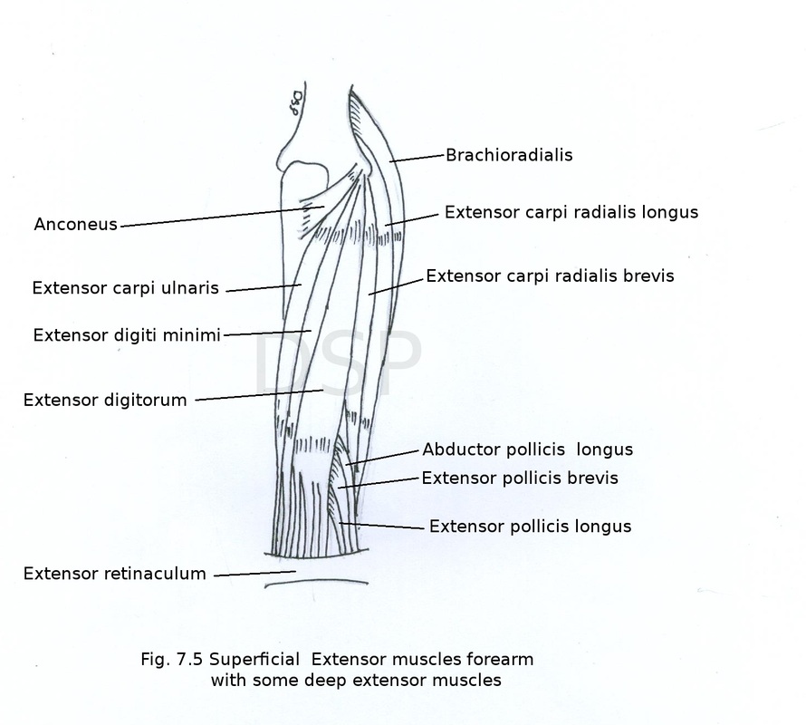
2. Posterior compartment of forearm
It is also extensor compartment of forearm.
Contents are extensor muscles, nerves and vessels.
Muscles are present in two groups superficial and deep.
Superficial muscles are 7
Origin : It shows origin from posterior surface of lateral epicondyle of humerus.
Insertion : It shows insertion on lateral surface of olecranon process and upper 1/4th part of posterior surface of shaft of ulna.
Nerve supply from radial nerve C7, C8, T1.
Action : It helps in extension at elbow joint.
2. Brachioradialis
Origin : It shows origin from upper two third part of lateral supracondylar ridge of humerus and lower part of lateral intermuscular septum.
Insertion : It shows insertion by a tendon near base of styloid process on lateral surface of radius.
Nerve supply : radial nerve C5, C6, C7, T1
Action : It helps in flexion at elbow joint during mid prone position.
3. Extensor carpi radialis longus
Origin : It shows origin from lower two third part of lateral supracondylar ridge, from common extensor origin ( lateral epicondyle) and from lower part of lateral intermuscular septum.
Insertion : It shows insertion on dorsal surface of second metacarpal bone near base.
Nerve supply from radial nerve C6, C7
Action : It helps in extension at wrist joint and abduction at wrist joint.
4. Extensor carpi radialis brevis
Origin : It shows origin from common extensor origin ( lateral epicondyle ) and from radial collateral ligament of elbow joint.
Insertion : It shows insertion on bases of second and third metacarpal bone on its dorsal surface.
Nerve supply : posterior interosseous nerve (the branch of the radial nerve C7, C8 )
Action : It helps in extension at wrist joint and abduction at wrist joint.
5. Extensor digitorum
Origin : common extensor origin (lateral epicondyle of humerus ) in lower part muscle divides into 4 tendons for the medial 4 fingers.
Insertion : With the help of dorsal digital expansion it shows insertion on the base of middle phalanx and base of distal phalanx.
Nerve supply : posterior interosseous nerve.
Action : It helps in extension at metacarpophalangeal and interphalangeal joints.
6. Extensor digiti minimi
Origin : It shows origin from common extensor origin ( lateral epicondyle humerus )
Insertion : Tendon of muscle join with extensor digitorum tendon of fifth finger on medial side ( dorsal digital expansion of that finger). Thus it shows insertion on base of middle phalanx and base of distal phalanx of fifth finger.
Nerve supply from posterior interosseous nerve C7, C8.
Action : Extension of fifth finger along with extensor tendon of extensor digitorum for fifth finger.
7. Extensor carpi ulnaris
Origin : It shows origin from common extensor origin (lateral epicondyle of humerus) and from posterior subcutaneous border of ulna along with origin of flexor carpi ulnaris with the help of an aponeurosis.
Insertion : It shows insertion on base of 5th metacarpal bone near its base.
Nerve supply : posterior interosseous nerve C7, C8.
Action : extension at wrist joint and adduction at wrist joint.
It is also extensor compartment of forearm.
Contents are extensor muscles, nerves and vessels.
Muscles are present in two groups superficial and deep.
Superficial muscles are 7
- Anconeus
- Brachioradialis
- Extensor carpi radialis longus
- Extensor carpi radialis brevis
- Extensor digitorum
- Extensor digiti minimi
- Extensor carpi ulnaris
Origin : It shows origin from posterior surface of lateral epicondyle of humerus.
Insertion : It shows insertion on lateral surface of olecranon process and upper 1/4th part of posterior surface of shaft of ulna.
Nerve supply from radial nerve C7, C8, T1.
Action : It helps in extension at elbow joint.
2. Brachioradialis
Origin : It shows origin from upper two third part of lateral supracondylar ridge of humerus and lower part of lateral intermuscular septum.
Insertion : It shows insertion by a tendon near base of styloid process on lateral surface of radius.
Nerve supply : radial nerve C5, C6, C7, T1
Action : It helps in flexion at elbow joint during mid prone position.
3. Extensor carpi radialis longus
Origin : It shows origin from lower two third part of lateral supracondylar ridge, from common extensor origin ( lateral epicondyle) and from lower part of lateral intermuscular septum.
Insertion : It shows insertion on dorsal surface of second metacarpal bone near base.
Nerve supply from radial nerve C6, C7
Action : It helps in extension at wrist joint and abduction at wrist joint.
4. Extensor carpi radialis brevis
Origin : It shows origin from common extensor origin ( lateral epicondyle ) and from radial collateral ligament of elbow joint.
Insertion : It shows insertion on bases of second and third metacarpal bone on its dorsal surface.
Nerve supply : posterior interosseous nerve (the branch of the radial nerve C7, C8 )
Action : It helps in extension at wrist joint and abduction at wrist joint.
5. Extensor digitorum
Origin : common extensor origin (lateral epicondyle of humerus ) in lower part muscle divides into 4 tendons for the medial 4 fingers.
Insertion : With the help of dorsal digital expansion it shows insertion on the base of middle phalanx and base of distal phalanx.
Nerve supply : posterior interosseous nerve.
Action : It helps in extension at metacarpophalangeal and interphalangeal joints.
6. Extensor digiti minimi
Origin : It shows origin from common extensor origin ( lateral epicondyle humerus )
Insertion : Tendon of muscle join with extensor digitorum tendon of fifth finger on medial side ( dorsal digital expansion of that finger). Thus it shows insertion on base of middle phalanx and base of distal phalanx of fifth finger.
Nerve supply from posterior interosseous nerve C7, C8.
Action : Extension of fifth finger along with extensor tendon of extensor digitorum for fifth finger.
7. Extensor carpi ulnaris
Origin : It shows origin from common extensor origin (lateral epicondyle of humerus) and from posterior subcutaneous border of ulna along with origin of flexor carpi ulnaris with the help of an aponeurosis.
Insertion : It shows insertion on base of 5th metacarpal bone near its base.
Nerve supply : posterior interosseous nerve C7, C8.
Action : extension at wrist joint and adduction at wrist joint.
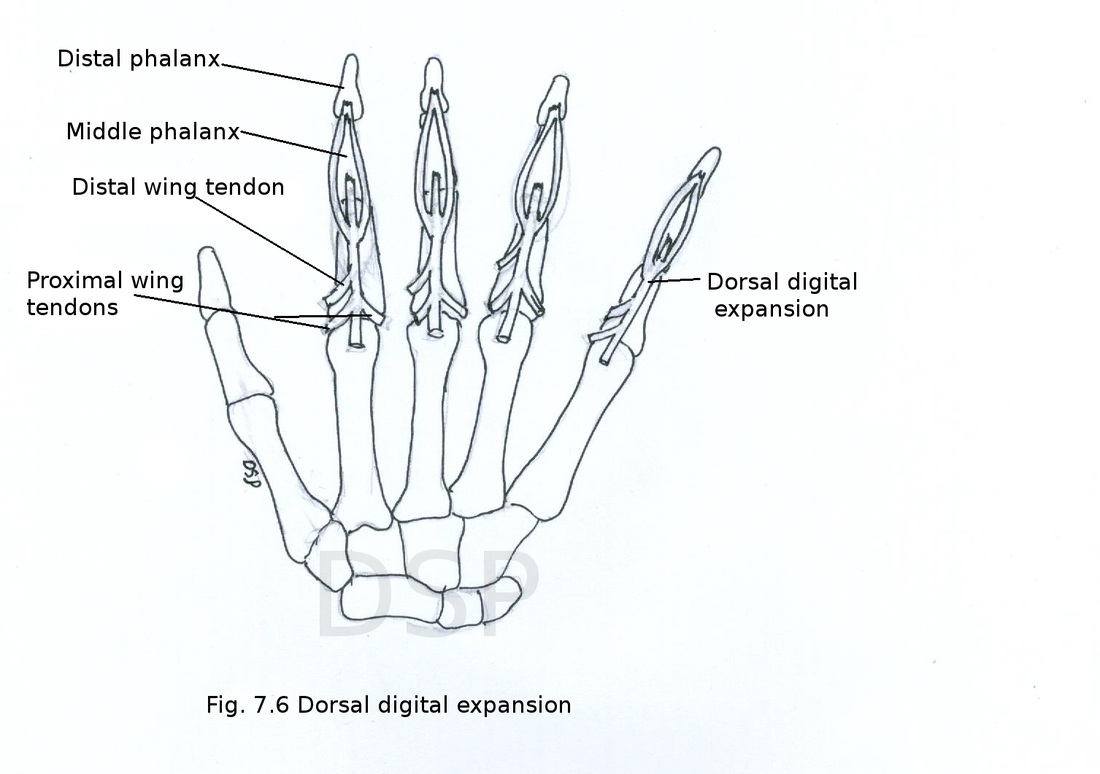
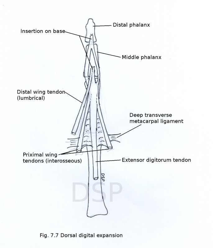
Dorsal digital expansion
It is a triangular expansion of extensor tendons of extensor digitorum on dorsal surface of metacarpal bones. It has base and apex. Base lies near metacarpophalangeal joint or metacarpal head and base is firmly attached with deep transverse metacarpal ligament. Apex near proximal interphalangeal joint divides into one median and two lateral slips. Median sleep shows insertion on base of middle phalanx and to lateral sleep unite and shows insertion on base of distal phalanx. Dorsal digital expansion shows attachment of tendons of lumbricals and interosseous muscles on lateral side and only interosseous muscle on medial side. Attached tendons are named as wing tendons proximal and distal wings tendons. Proximal wing tendons are formed by palmar and dorsal interossei which join with dorsal digital expansion near metacarpophalangeal joint and distal wing tendons are formed by tendons of lumbricals which joins with dorsal digital expansion near proximal interphalangeal joints.
It is a triangular expansion of extensor tendons of extensor digitorum on dorsal surface of metacarpal bones. It has base and apex. Base lies near metacarpophalangeal joint or metacarpal head and base is firmly attached with deep transverse metacarpal ligament. Apex near proximal interphalangeal joint divides into one median and two lateral slips. Median sleep shows insertion on base of middle phalanx and to lateral sleep unite and shows insertion on base of distal phalanx. Dorsal digital expansion shows attachment of tendons of lumbricals and interosseous muscles on lateral side and only interosseous muscle on medial side. Attached tendons are named as wing tendons proximal and distal wings tendons. Proximal wing tendons are formed by palmar and dorsal interossei which join with dorsal digital expansion near metacarpophalangeal joint and distal wing tendons are formed by tendons of lumbricals which joins with dorsal digital expansion near proximal interphalangeal joints.
Deep group of extensor muscles of forearm
These are five in number
Origin : It shows origin of superficial fibres from lateral epicondyle, radial collateral ligament, annular ligament and deep fibres from supinator crest of ulna also from triangular area anterior to supinator crest.
Insertion : Superficial fibres shows insertion on upper one third part of lateral surface of radius anterior part, deep fibres shows insertion on radius between anterior and posterior oblique lines but direction of fibre is horizontal.
Nerve supply : posterior interosseous nerve C6, C7.
Action : supination of forearm.
2. Abductor pollicis longus
Origin : It shows origin from posterior surface of radius and ulna upper part and upper part of interosseous membrane.
Insertion : Its tendon shows insertion on radial side of base of first metacarpal bone.
Nerve supply from posterior interosseous nerve.
Action : It helps in abduction and extension of thumb at carpometacarpal joint.
3. Extensor pollicis brevis
Origin : It shows origin from posterior surface of radius below origin of abductor pollicis longus and part of interosseous membrane.
Insertion : It shows insertion on base of proximal phalanx of thumb on its dorsal surface.
Nerve supply : posterior interosseous nerve C7, C8.
Action : extension of proximal phalanx of thumb.
4. Extensor pollicis longus
Origin : It shows origin from posterior surface of shaft of ulna below origin of abductor pollicis longus and also part of interosseous membrane in relation with that part of ulna.
Insertion : It shows insertion by a tendon on base of distal phalanx of thumb on dorsal surface
Nerve supply : posterior interosseous nerve C7, C8.
Action : extension of distal phalanx of thumb.
5. Extensor indicis
Origin : Its shows origin from posterior surface of a ulna below origin of extensor pollicis longus and part of interosseous membrane in relation to that part of ulna.
Insertion : tendon of extensor indicis join with tendon of extensor digitorum for index finger on ulnar side.
Nerve supply : posterior interosseous nerve.
Action : It will help in extension of index finger.
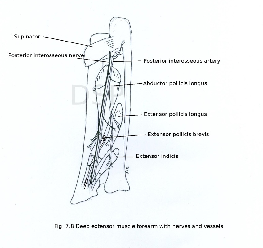
Posterior interosseous nerve (deep terminal branch of the radial nerve)
It is a terminal branch of radial nerve. Which runs in extensor compartment of forearm. This branch arises from radial nerve in the cubital fossa anterior to lateral epicondyle of humerus. It goes to extensor compartment of forearm by passing between superficial and deep fibres of supinator muscle. In extensor compartment it lies between superficial and deep group of extensor muscles after supplying extensor muscles it goes down on posterior surface of interosseous membrane. It shows a pseudoganglion and finally gives branches to wrist joint and carpal joints .
Branches
1. Muscular branches
It gives muscular branches to extensor carpi radialis brevis, supinator, extensor digitorum, extensor digiti minimi, extensor carpi ulnaris, extensor pollicis longus, extensor indicis, abductor pollicis longus and extensor pollicis brevis.
2. Articular branches
It gives articular branches to wrist joint, inferior radioulnar joint, intercarpal joint and inter metacarpal joints.
3. Sensory branches
It gives sensory branches to interosseous membrane, radius and ulna.
Posterior interosseous artery
It is the branch of common interosseous artery (branch of ulnar artery). It enters in posterior compartment between upper border of interosseous membrane and oblique cord. Then it runs on posterior surface of interosseous membrane along with posterior interosseous nerve. It goes down between superficial and deep groups of extensor muscles of forearm. It anastomoses with anterior interosseous artery in lower part. It also gives an interosseous recurrent artery which forms anastomosis on posterior part of lateral epicondyle of humerus by joining with middle collateral branch of profunda brachii, posterior ulnar recurrent and ulnar collateral artery.
These are five in number
- Supinator
- Abductor pollicis longus
- Extensor pollicis brevis
- Extensor pollicis longus
- Extensor indicis
Origin : It shows origin of superficial fibres from lateral epicondyle, radial collateral ligament, annular ligament and deep fibres from supinator crest of ulna also from triangular area anterior to supinator crest.
Insertion : Superficial fibres shows insertion on upper one third part of lateral surface of radius anterior part, deep fibres shows insertion on radius between anterior and posterior oblique lines but direction of fibre is horizontal.
Nerve supply : posterior interosseous nerve C6, C7.
Action : supination of forearm.
2. Abductor pollicis longus
Origin : It shows origin from posterior surface of radius and ulna upper part and upper part of interosseous membrane.
Insertion : Its tendon shows insertion on radial side of base of first metacarpal bone.
Nerve supply from posterior interosseous nerve.
Action : It helps in abduction and extension of thumb at carpometacarpal joint.
3. Extensor pollicis brevis
Origin : It shows origin from posterior surface of radius below origin of abductor pollicis longus and part of interosseous membrane.
Insertion : It shows insertion on base of proximal phalanx of thumb on its dorsal surface.
Nerve supply : posterior interosseous nerve C7, C8.
Action : extension of proximal phalanx of thumb.
4. Extensor pollicis longus
Origin : It shows origin from posterior surface of shaft of ulna below origin of abductor pollicis longus and also part of interosseous membrane in relation with that part of ulna.
Insertion : It shows insertion by a tendon on base of distal phalanx of thumb on dorsal surface
Nerve supply : posterior interosseous nerve C7, C8.
Action : extension of distal phalanx of thumb.
5. Extensor indicis
Origin : Its shows origin from posterior surface of a ulna below origin of extensor pollicis longus and part of interosseous membrane in relation to that part of ulna.
Insertion : tendon of extensor indicis join with tendon of extensor digitorum for index finger on ulnar side.
Nerve supply : posterior interosseous nerve.
Action : It will help in extension of index finger.
Posterior interosseous nerve (deep terminal branch of the radial nerve)
It is a terminal branch of radial nerve. Which runs in extensor compartment of forearm. This branch arises from radial nerve in the cubital fossa anterior to lateral epicondyle of humerus. It goes to extensor compartment of forearm by passing between superficial and deep fibres of supinator muscle. In extensor compartment it lies between superficial and deep group of extensor muscles after supplying extensor muscles it goes down on posterior surface of interosseous membrane. It shows a pseudoganglion and finally gives branches to wrist joint and carpal joints .
Branches
1. Muscular branches
It gives muscular branches to extensor carpi radialis brevis, supinator, extensor digitorum, extensor digiti minimi, extensor carpi ulnaris, extensor pollicis longus, extensor indicis, abductor pollicis longus and extensor pollicis brevis.
2. Articular branches
It gives articular branches to wrist joint, inferior radioulnar joint, intercarpal joint and inter metacarpal joints.
3. Sensory branches
It gives sensory branches to interosseous membrane, radius and ulna.
Posterior interosseous artery
It is the branch of common interosseous artery (branch of ulnar artery). It enters in posterior compartment between upper border of interosseous membrane and oblique cord. Then it runs on posterior surface of interosseous membrane along with posterior interosseous nerve. It goes down between superficial and deep groups of extensor muscles of forearm. It anastomoses with anterior interosseous artery in lower part. It also gives an interosseous recurrent artery which forms anastomosis on posterior part of lateral epicondyle of humerus by joining with middle collateral branch of profunda brachii, posterior ulnar recurrent and ulnar collateral artery.
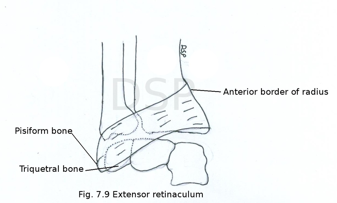
Extensor retinaculum
It is an oblique fibrous band present on back of wrist joint directed downwards and medially. It develops from deep fascia and about 2 centimetre broad. This is for holding extensor tendon in place on dorsum of wrist.
Attachments
Medially : on triquetral and pisiform bone
Laterally : anterior border of radius in lower part
Structures deep to retinaculum
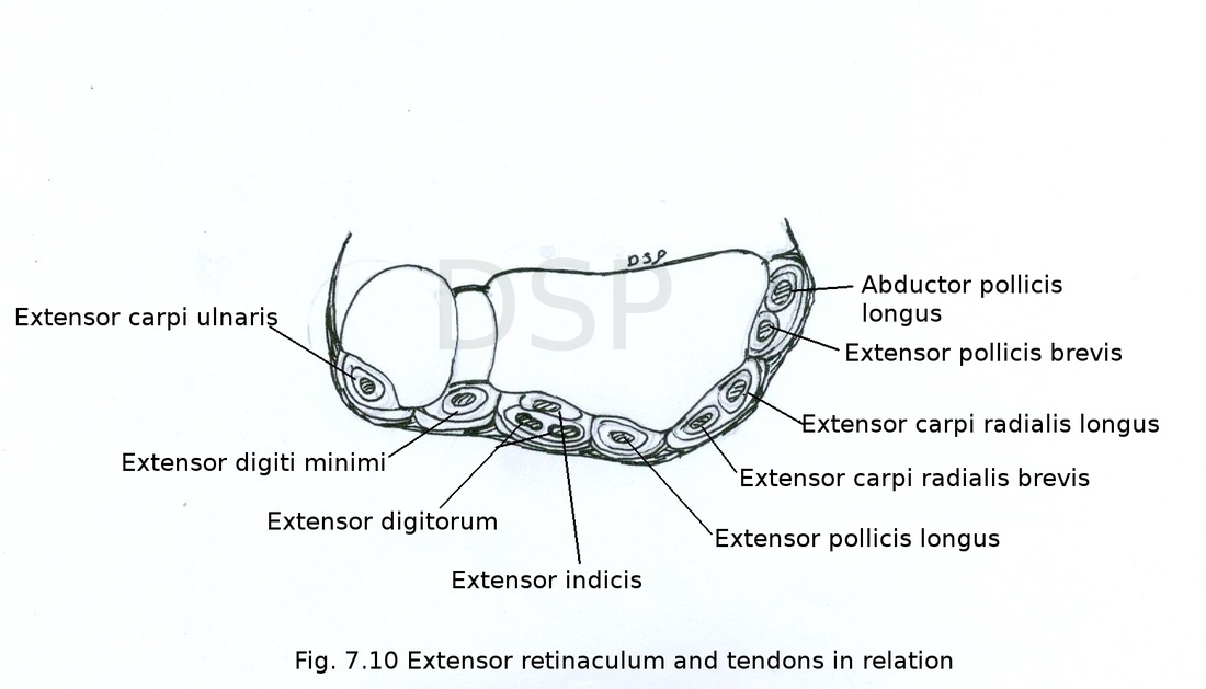
Retinaculum gives fibrous septum and divides into 6 compartments.
Structures passing through compartments from lateral to medial side
It is an oblique fibrous band present on back of wrist joint directed downwards and medially. It develops from deep fascia and about 2 centimetre broad. This is for holding extensor tendon in place on dorsum of wrist.
Attachments
Medially : on triquetral and pisiform bone
Laterally : anterior border of radius in lower part
Structures deep to retinaculum
Retinaculum gives fibrous septum and divides into 6 compartments.
Structures passing through compartments from lateral to medial side
- Abductor pollicis longus, Extensor pollicis brevis
- Extensor carpi radialis longus, Extensor carpi radialis brevis
- Extensor pollicis longus
- Four tendons of Extensor digitorum, Extensor indicis, Posterior interosseous nerve and Anterior interosseous artery
- Extensor digiti minimi
- Extensor carpi ulnaris