BONES THORAX
RIBS
Ribs are elongated plate like flat bones. there are 12 pairs of ribs. These are obliquely placed showing articulation posteriorly with thoracic vertebra and anteriorly with sternum but last two ribs articulate with sternum with the help of costal cartilages.
Types of ribs
True rib : Upper seven pairs of ribs are directly articulates with sternum through their cartilages called true ribs.
False rib : 8 to 12th ribs are false ribs. 8th, 9th and 10th ribs are called vertebro-chondral ribs because they articulate with sternum indirectly by joining their upper ends of costal cartilages. 11th and 12th ribs are floating ribs because their lateral ends are free.
Typical and atypical ribs : 3rd to 9th ribs are typical ribs because they show similar features and 1st, 2nd, 10th, 11th and 12th ribs are atypical or Peculiar because they are having different identification features.
Other features of ribs : Length of ribs gradually increases from first rib to seventh rib and then length decreases up to 12th rib. ribs goes obliquely downward and forward. So Seventh rib is having maximum length and maximum obliquity is present in 9th rib.
Typical ribs :
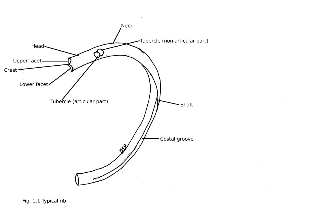
3rd to 9th ribs are typical ribs. Typical ribs shows two ends anterior (sternal) and posterior (vertebral) and shaft. Anterior end is slightly lower than posterior end and it shows concave depression for articulation with its costal cartilage. Posterior end shows head, neck and tubercle. Head shows two facets separated by crest. Lower facet is large and shows articulation with body of corresponding vertebra. Upper facet is small in size and shows articulation with body of upper vertebra. Crest shows attachment with intervertebral disc by a ligament. Neck is flat part of rib goes postero-laterally from head to tubercle of rib anterior to transverse process. It shows two surfaces anterior and posterior to borders superior and inferior. Anterior surface is smooth and posterior surface rough. Superior border is thin like a crest and inferior border is rounded. Tubercle is a small elevation present on the outer surface of rib at the junction of neck and shaft. It shows medial articular part and lateral non articular part. Medial articular part shows articulation with costal facet present on transverse process of corresponding vertebra. Shaft is flat and plate like part of rib. Shaft goes first backward and laterally then changes direction and goes forward forming angle of rib. It shows two surfaces outer and inner and to borders upper and lower. Outer surface shows a line which goes downward and laterally. Inner surface shows smooth surface lined by pleura. Inner surface shows a costal groove in relation with its lower part. Costal groove shows upper and lower lips. Costal groove contains posterior intercostal vessels and intercostal nerve. Upper border of shaft is broad shows outer and inner lips while lower border of shaft is thin and sharp.
Attachments:
Crest of rib shows attachment of intra-articular ligament connected with inter-vertebral disc. Head shows attachment of radiate ligament anteriorly. Neck is covered by costal pleura anteriorly. Posterior surface of neck shows attachment of costotransverse ligament. Superior border of neck shows attachment of superior costotransverse ligament. Inferior border of neck shows attachment of intercostal membrane of lower intercostal space. Non articular part of tubercle shows attachment of lateral costotransverse ligament. Shaft near angle on outer surface shows attachment of posterior layer of thoracolumbar fascia and lateral fibres of sacrospinalis muscle and medially levator costae with sacrospinalis. Near sternal end there is an oblique line. Anterior end shows origin of external oblique and serratus anterior (in upper eight ribs) while origin of external oblique and latissimus dorsi (9, 10 and 11th ribs) separated by this line. Upper lip of costal groove shows origin of intercostalis intimus in its middle part and floor between two lips shows origin of internal intercostal muscle. Outer lip of upper border shows insertion of external intercostal muscle and inner lip shows attachment of internal intercostal and intercostalis intimus muscles. Lower border of shaft shows origin of external intercostal muscle.
Atypical ribs :
1st, 2nd, 10th, 11th and 12th ribs are atypical.
First rib
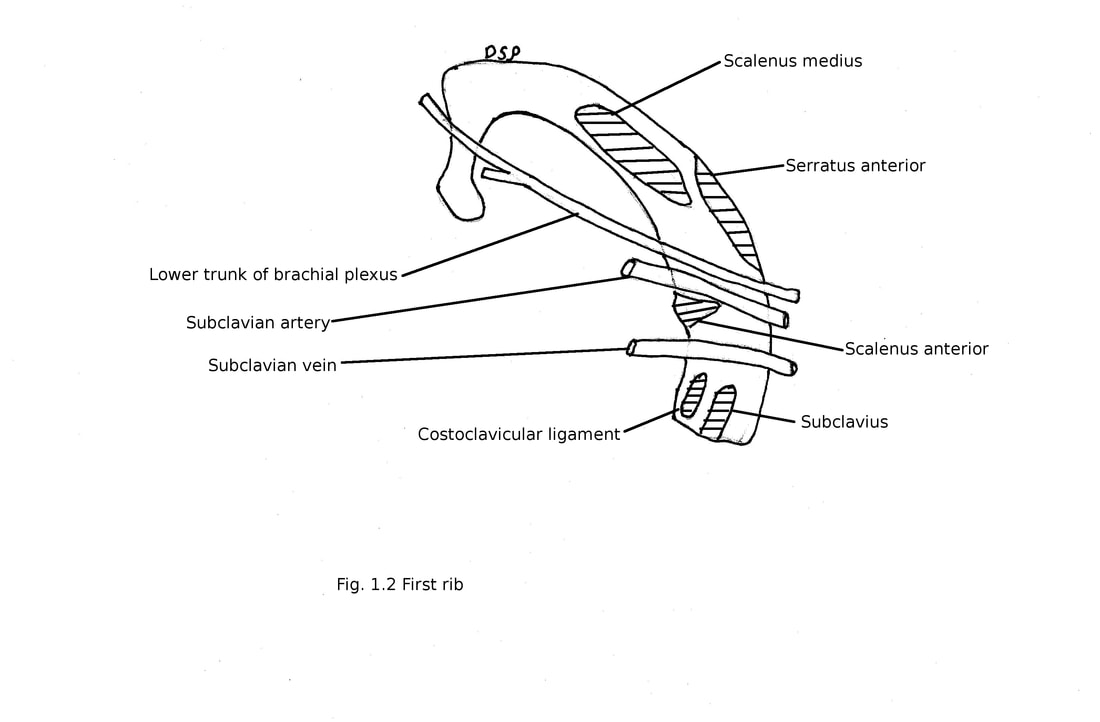
It is short, flat and strong of among all true ribs. It shows two ends anterior (sternal) and posterior (vertebral) and shaft. Anterior end is 3-4 cm lower than posterior end and continue with first costal cartilage forms first chondrosternal joint. Posterior end shows head, neck and tubercle. Head is small with round articular facet for articulation with body of first thoracic vertebra. Neck is rounded goes postero-laterally. Tubercle is big articulating with transverse process of first thoracic vertebra. Shaft is flat shows two surfaces upper and lower with two borders outer and inner. Upper surface shows two oblique grooves and a scalene tubercle near inner border. The grooves are for subclavian vein (anterior part), subclavian artery and lower trunk of brachial plexus (posterior part). Lower surface is smooth without costal groove. Inner border is concave in shape. Outer border is convex and thick posterior part and thin anteriorly.
Side determination of first rib : Place rib on a flat surface anterior and posterior end touch surface it indicate side of that rib. But if wrongly placed posterior end will be lifted.
Attachments:
Anterior end shows attachment of subclavius muscle and attachment of costoclavicular ligament. Scalene tubercle show insertion of scalenus anterior muscle. Scalenus medius shows insertion behind groove. Origin of first digitation of serratus anterior present behind groove also present. Suprapleural membrane shows attachment on inner border
Second rib
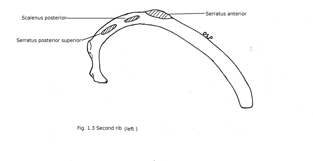
It is same like first rib but length of it is double that of first rib. Nonarticular part of tubercle is small. Shaft shows outer and inner surface. Outer surface of shaft is convex facing upward and outward. A tuberosity is present in the middle of shaft. Inner surface faces downward and medially.
Attachments:
Tubercle shows origin of part of first and second digitation of serratus anterior. Upper border shows insertion of scalenus posterior.
Tenth rib
It shows all features of a typical rib but shows a single facet to articulate with body of tenth thoracic vertebra.
Eleventh rib and Twelfth rib
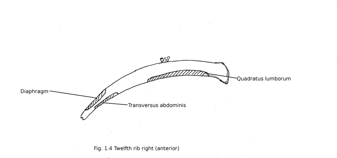
These are short ribs. Free ends are caped by cartilage. Eleventh rib shows a single large facet on head for articulation with body of eleventh thoracic vertebra. It has no neck , no tubercle. Twelfth rib a single facet on its head for articulation with body of twelfth thoracic vertebra. It is without neck, tubercle, angle and costal groove. Twelfth rib shows anterior and posterior surface, upper and lower borders.
Attachments:
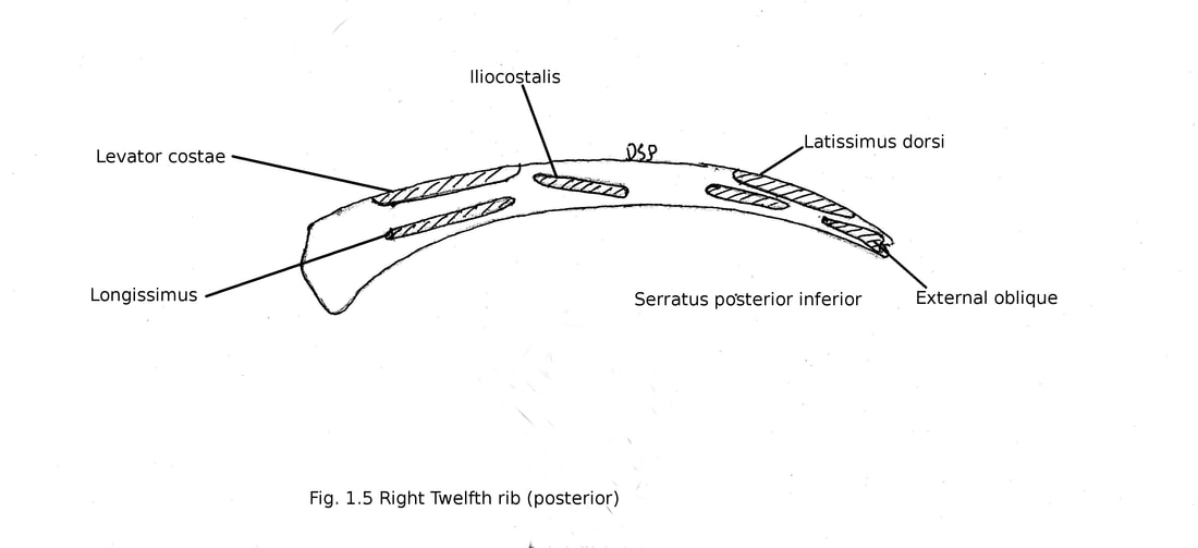
Twelfth rib on inner surface shows insertion of quadratus lumborum on medial half with fascia covering it, insertion of internal intercostal muscle on upper border, origin of diaphragm, transversus abdominis just below diaphragm. Costodiaphragmatic recess comes in relation with medial three forth part of twelfth rib.
Twelfth rib on outer surface shows attachment of costotransverse ligament, lumbocostal ligament, insertion of lowest levator costae, iliocostalis and longissimus (medial part), insertion of serratus posterior inferior, origin of external oblique and latissimus dorsi (lateral part).
While lower border of twelfth rib show attachment of lumbocostal ligament, middle layer of thoracolumbar fascia, lateral arcuate ligament.
Ossification:
A typical rib ossifies from one primary centre and three secondary centres. Primary centre appear in shaft 8th weeks of intrauterine life. The secondary centres one for articular part, one for non articular part of tubercle and one for head appears during 12-15 years and fuses with shaft after 20th years.
First rib shows one primary centre for shaft and two secondary centres for head and tubercle.
Eleventh and twelfth rib shows one primary centre for shaft and one secondary centres for head.
Applied anatomy:
1) Cervical rib : A rib which develops from transverse processes of seventh cervical vertebra. It may cause thoracic inlet syndrome by pressing lower trunk of brachial plexus and subclavian artery because of a fibrous band connecting anterior part of first rib and cervical rib.
2) Fracture of ribs : It is common in compression injuries of chest. Ribs get fractured in front of angle (weakest part of rib). Fracture causes injury to pleura. First and last two ribs are not fractured.
3) Flail chest : When ribs get fractured at two or more points that segment of ribs go in and out during each respiratory movement.
4) Lumbar rib : A rib which develops from transverse processes of first lumbar vertebra.
COSTAL CARTILAGES
These are hyaline cartilages present on anterior aspect of ribs. It provides elasticity to thoracic wall. First to seven costal cartilages anteriorly shows attachment directly with sternum. Eight to ten cartilage join with each other forming costal margin. Eleven and twelfth cartilage are present on the tip of respective ribs. Each costal cartilage shows two surfaces anterior and posterior, two borders upper and lower. Medially first cartilage shows formation of primary cartilaginous joint with manubrium. Second to seventh cartilages shows formation of synovial joints with sternum.
Attachments
Anterior surface of first cartilage shows attachment of sternoclavicular disc, joint capsule of sternoclavicular joint, sternoclavicular ligament. costoclvicular ligament and subclavius shows attachment on first costal cartilage. Anterior surface of second to seventh cartilage shows origin of pectoralis major muscle. Muscles of abdomen internal oblique and rectus abdominis show attachment on anterior surfaces of seventh to ninth and fifth to seventh cartilage.
Posterior surface of first cartilage shows origin of sternothyroid. Posterior surfaces of second to sixth cartilage shows insertion of sternocostalis. Posterior surfaces of seventh to twelfth cartilage shows attachment of diaphragm and transversus abdominis.
Superior and inferior borders of cartilages shows attachment of external and internal intercostal muscles.
These are short ribs. Free ends are caped by cartilage. Eleventh rib shows a single large facet on head for articulation with body of eleventh thoracic vertebra. It has no neck , no tubercle. Twelfth rib a single facet on its head for articulation with body of twelfth thoracic vertebra. It is without neck, tubercle, angle and costal groove. Twelfth rib shows anterior and posterior surface, upper and lower borders.
Attachments:
Twelfth rib on inner surface shows insertion of quadratus lumborum on medial half with fascia covering it, insertion of internal intercostal muscle on upper border, origin of diaphragm, transversus abdominis just below diaphragm. Costodiaphragmatic recess comes in relation with medial three forth part of twelfth rib.
Twelfth rib on outer surface shows attachment of costotransverse ligament, lumbocostal ligament, insertion of lowest levator costae, iliocostalis and longissimus (medial part), insertion of serratus posterior inferior, origin of external oblique and latissimus dorsi (lateral part).
While lower border of twelfth rib show attachment of lumbocostal ligament, middle layer of thoracolumbar fascia, lateral arcuate ligament.
Ossification:
A typical rib ossifies from one primary centre and three secondary centres. Primary centre appear in shaft 8th weeks of intrauterine life. The secondary centres one for articular part, one for non articular part of tubercle and one for head appears during 12-15 years and fuses with shaft after 20th years.
First rib shows one primary centre for shaft and two secondary centres for head and tubercle.
Eleventh and twelfth rib shows one primary centre for shaft and one secondary centres for head.
Applied anatomy:
1) Cervical rib : A rib which develops from transverse processes of seventh cervical vertebra. It may cause thoracic inlet syndrome by pressing lower trunk of brachial plexus and subclavian artery because of a fibrous band connecting anterior part of first rib and cervical rib.
2) Fracture of ribs : It is common in compression injuries of chest. Ribs get fractured in front of angle (weakest part of rib). Fracture causes injury to pleura. First and last two ribs are not fractured.
3) Flail chest : When ribs get fractured at two or more points that segment of ribs go in and out during each respiratory movement.
4) Lumbar rib : A rib which develops from transverse processes of first lumbar vertebra.
COSTAL CARTILAGES
These are hyaline cartilages present on anterior aspect of ribs. It provides elasticity to thoracic wall. First to seven costal cartilages anteriorly shows attachment directly with sternum. Eight to ten cartilage join with each other forming costal margin. Eleven and twelfth cartilage are present on the tip of respective ribs. Each costal cartilage shows two surfaces anterior and posterior, two borders upper and lower. Medially first cartilage shows formation of primary cartilaginous joint with manubrium. Second to seventh cartilages shows formation of synovial joints with sternum.
Attachments
Anterior surface of first cartilage shows attachment of sternoclavicular disc, joint capsule of sternoclavicular joint, sternoclavicular ligament. costoclvicular ligament and subclavius shows attachment on first costal cartilage. Anterior surface of second to seventh cartilage shows origin of pectoralis major muscle. Muscles of abdomen internal oblique and rectus abdominis show attachment on anterior surfaces of seventh to ninth and fifth to seventh cartilage.
Posterior surface of first cartilage shows origin of sternothyroid. Posterior surfaces of second to sixth cartilage shows insertion of sternocostalis. Posterior surfaces of seventh to twelfth cartilage shows attachment of diaphragm and transversus abdominis.
Superior and inferior borders of cartilages shows attachment of external and internal intercostal muscles.
THE STERNUM
Sternum is short flat sword like bone. It shows three parts manubrium, body and xiphoid processes.
Manubrium : It is quadrilateral in shape. Vertebral level of it is third and forth thoracic vertebra. It shows two surfaces anterior and posterior, four borders upper, lower and two lateral. Anterior surface shows concavity from above downwards and convex from side to side. Posterior surface is concave. Upper border shows supra-sternal notch of jugular notch in the middle and clavicular notch on each side. Medial end of clavicle articulates with clavicular notch forming sternoclavicular joint. Lower border shows articulation with upper part of body of sternum forming manubriosternal joint (secondary cartilaginous joint). An forward projecting sternal angle (angle of louis) forms at manubriosternal joint. Two lateral borders forms joint with first costal cartilage (primary cartilaginous joint) and in lower part shows half facet for articulation with second costal cartilage.
Body of sternum : It is long narrow and thin part. It lies opposite fifth to ninth thoracic vertebra. It shows broadest part at the level of fifth costal cartilage. It shows two ends (upper and lower), two lateral borders and two surfaces (anterior and posterior). Upper end forms a secondary cartilaginous joint with manubrium forming manubriosternal joint. Sternal angle forms here. Lower border of fourth thoracic vertebra lies at this level. Lower end forms joint with xiphoid process. Lateral borders shows four complete facets for third to sixth costal cartilages and half facets for second and seventh cartilage. Anterior surface is flat. It shows three transverse ridges which indicate body is formed by four segments known as sternebrae. Posterior surface is concave with ill defined three transverse ridges.
Xiphoid process : It is smallest part present on lower aspect of body. Its shape may be triangular pointed, bifid, perforated, deflected to one side. Upper end joins with body and forms xiphisternal joint.
Attachments
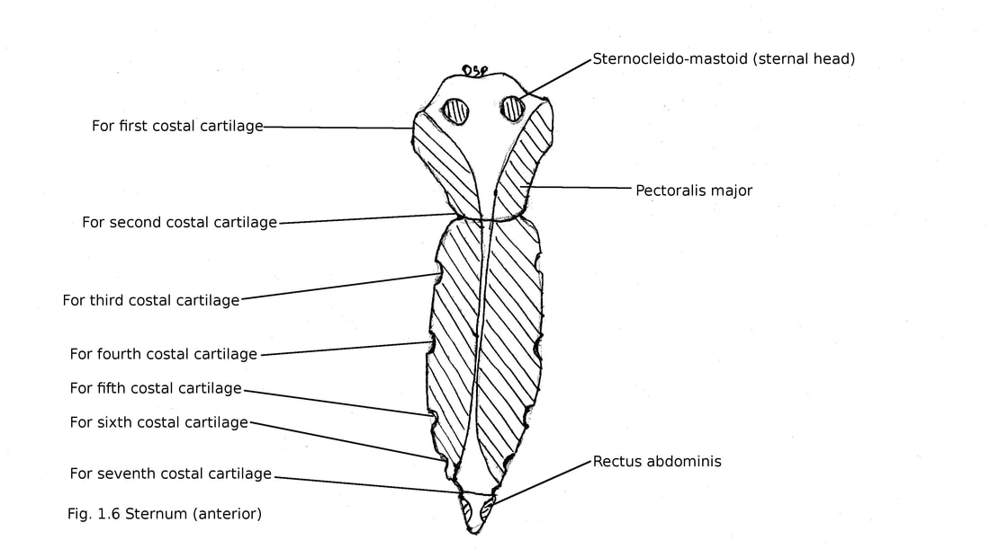
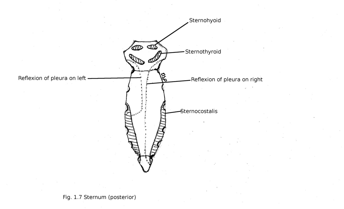
Anterior surface of manubrium shows origin of pectoralis major, sternal head of sternocleidomastoid. Posterior surface of manubrium shows origin of sternohyoid, sternothyroid. Supra-sternal notch shows attachment of interclavicular ligament and investing layer of deep cervical fascia. Lateral margins shows attachment of capsule of sternoclavicular joint.
Anterior surface of body of sternum shows origin of pectoralis major. Posterior surface shows origin of sternocostalis near lower part.
Anterior surface xiphoid process shows insertion of rectus abdominis, aponeuroses of external and internal oblique muscles. Posterior surface of xiphoid process shows origin of diaphragm. Lateral margins shows attachment of aponeuroses of internal oblique and transversus abdominis muscles. Lower end shows attachment of linea alba.
Ossification :
It develops from five centres of ossification. One centre for manubrium appears during fifth month of intrauterine life. Remaining four centres forms four sternebrae. First and second sternebrae appears in fifth month of intrauterine life, third and fourth sternebrae are paired appear in fifth and sixth month or seventh and eight month of intrauterine life. All these fuse with each other at 25th year. Xiphoid process appear in third year after birth.
Sternum is short flat sword like bone. It shows three parts manubrium, body and xiphoid processes.
Manubrium : It is quadrilateral in shape. Vertebral level of it is third and forth thoracic vertebra. It shows two surfaces anterior and posterior, four borders upper, lower and two lateral. Anterior surface shows concavity from above downwards and convex from side to side. Posterior surface is concave. Upper border shows supra-sternal notch of jugular notch in the middle and clavicular notch on each side. Medial end of clavicle articulates with clavicular notch forming sternoclavicular joint. Lower border shows articulation with upper part of body of sternum forming manubriosternal joint (secondary cartilaginous joint). An forward projecting sternal angle (angle of louis) forms at manubriosternal joint. Two lateral borders forms joint with first costal cartilage (primary cartilaginous joint) and in lower part shows half facet for articulation with second costal cartilage.
Body of sternum : It is long narrow and thin part. It lies opposite fifth to ninth thoracic vertebra. It shows broadest part at the level of fifth costal cartilage. It shows two ends (upper and lower), two lateral borders and two surfaces (anterior and posterior). Upper end forms a secondary cartilaginous joint with manubrium forming manubriosternal joint. Sternal angle forms here. Lower border of fourth thoracic vertebra lies at this level. Lower end forms joint with xiphoid process. Lateral borders shows four complete facets for third to sixth costal cartilages and half facets for second and seventh cartilage. Anterior surface is flat. It shows three transverse ridges which indicate body is formed by four segments known as sternebrae. Posterior surface is concave with ill defined three transverse ridges.
Xiphoid process : It is smallest part present on lower aspect of body. Its shape may be triangular pointed, bifid, perforated, deflected to one side. Upper end joins with body and forms xiphisternal joint.
Attachments
Anterior surface of manubrium shows origin of pectoralis major, sternal head of sternocleidomastoid. Posterior surface of manubrium shows origin of sternohyoid, sternothyroid. Supra-sternal notch shows attachment of interclavicular ligament and investing layer of deep cervical fascia. Lateral margins shows attachment of capsule of sternoclavicular joint.
Anterior surface of body of sternum shows origin of pectoralis major. Posterior surface shows origin of sternocostalis near lower part.
Anterior surface xiphoid process shows insertion of rectus abdominis, aponeuroses of external and internal oblique muscles. Posterior surface of xiphoid process shows origin of diaphragm. Lateral margins shows attachment of aponeuroses of internal oblique and transversus abdominis muscles. Lower end shows attachment of linea alba.
Ossification :
It develops from five centres of ossification. One centre for manubrium appears during fifth month of intrauterine life. Remaining four centres forms four sternebrae. First and second sternebrae appears in fifth month of intrauterine life, third and fourth sternebrae are paired appear in fifth and sixth month or seventh and eight month of intrauterine life. All these fuse with each other at 25th year. Xiphoid process appear in third year after birth.
VERTEBRAL COLUMN OF THORAX
Thoracic part of vertebral column shows curvatures concave anteriorly and concave laterally towards left side.
Thoracic region shows twelve thoracic vertebra. Thoracic vertebra are typical and atypical.
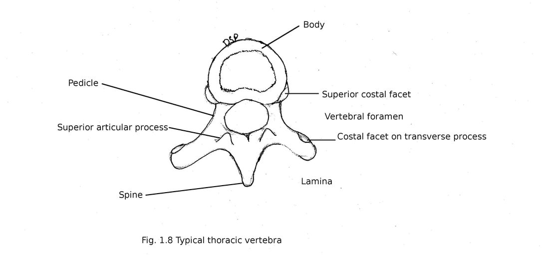
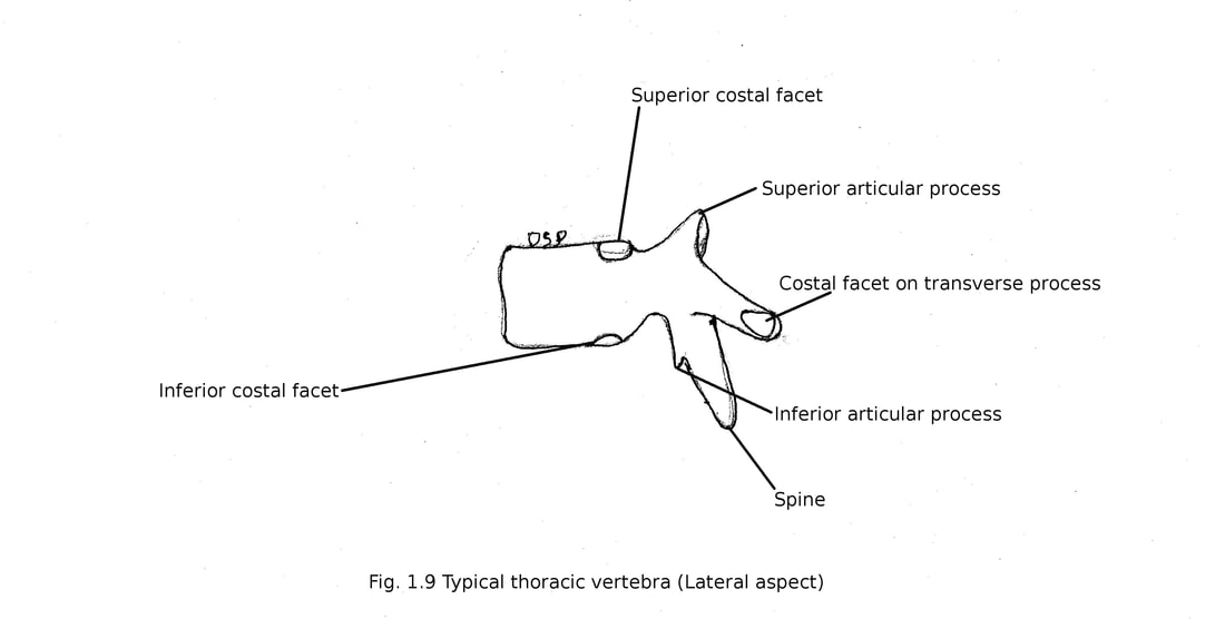
Typical thoracic vertebra : Second to eight thoracic vertebra are typical. Body of vertebra heart shaped. Transverse and antero-posterior diameters are same. It shows two costal facets on bodies on both sides near margins. Upper facet slightly larger than lower facets. Upper facets articulate with head of corresponding rib while lower facet articulate with head of lower rib. Vertebral foramens are small and circular in size. Pedicles are short and goes posteriorly. Superior vertebral notch is not well defined and inferior vertebral notch deep and prominent. Lamina are thick and short overlapping each other from above downward. Superior articular processes goes upward and facets are flat facing slightly upward and postero-laterally. Inferior articular processes get fused with lamina. Inferior articular facets faces slightly downward and anteriorly-medially. Transverse processes are large goes laterally backward from junction of pedicle and lamina. Near tip of transverse precess shows costal facets for articulation with tubercle of corresponding ribs. Facets on transverse process are concave in upper six thoracic vertebra facing antero-laterally but in other it is flat facing supero-laterally and upward. Spine is long goes downward and posteriorly. Fifth to ninth thoracic spines are long and more oblique.
Attachments :
Body shows attachment of anterior and posterior longitudinal ligaments near margins anteriorly and posteriorly. Transverse processes shows attachment of lateral costo-transverse ligament near tip, superior costo-transverse ligament near lower margin, inferior costo-transverse ligament on anterior surface, inter transverse muscles near upper and lower margins and levator costae on posterior surface. Upper and lower margins of laminae on anterior aspect shows attachment of ligamenta flava. Spine shows attachment of interspinous, supraspinous ligaments, trapezius, rhomboids major and minor, latissimus dorsi, serratus posterior superior, serratus posterior inferior, medial group of erector spinae muscles.
Thoracic part of vertebral column shows curvatures concave anteriorly and concave laterally towards left side.
Thoracic region shows twelve thoracic vertebra. Thoracic vertebra are typical and atypical.
Typical thoracic vertebra : Second to eight thoracic vertebra are typical. Body of vertebra heart shaped. Transverse and antero-posterior diameters are same. It shows two costal facets on bodies on both sides near margins. Upper facet slightly larger than lower facets. Upper facets articulate with head of corresponding rib while lower facet articulate with head of lower rib. Vertebral foramens are small and circular in size. Pedicles are short and goes posteriorly. Superior vertebral notch is not well defined and inferior vertebral notch deep and prominent. Lamina are thick and short overlapping each other from above downward. Superior articular processes goes upward and facets are flat facing slightly upward and postero-laterally. Inferior articular processes get fused with lamina. Inferior articular facets faces slightly downward and anteriorly-medially. Transverse processes are large goes laterally backward from junction of pedicle and lamina. Near tip of transverse precess shows costal facets for articulation with tubercle of corresponding ribs. Facets on transverse process are concave in upper six thoracic vertebra facing antero-laterally but in other it is flat facing supero-laterally and upward. Spine is long goes downward and posteriorly. Fifth to ninth thoracic spines are long and more oblique.
Attachments :
Body shows attachment of anterior and posterior longitudinal ligaments near margins anteriorly and posteriorly. Transverse processes shows attachment of lateral costo-transverse ligament near tip, superior costo-transverse ligament near lower margin, inferior costo-transverse ligament on anterior surface, inter transverse muscles near upper and lower margins and levator costae on posterior surface. Upper and lower margins of laminae on anterior aspect shows attachment of ligamenta flava. Spine shows attachment of interspinous, supraspinous ligaments, trapezius, rhomboids major and minor, latissimus dorsi, serratus posterior superior, serratus posterior inferior, medial group of erector spinae muscles.
Atypical thoracic vertebra : First, ninth, tenth, eleventh, twelfth are atypical thoracic vertebra.
First thoracic vertebra
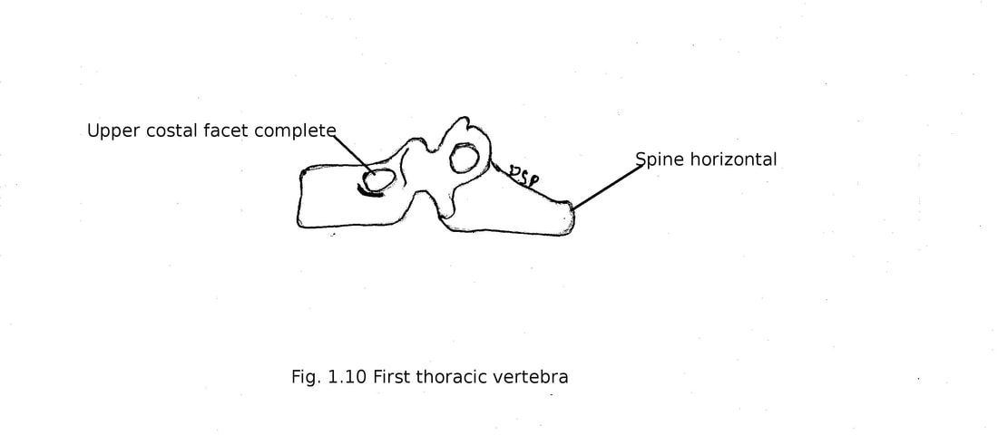
Body of it is broad like a cervical vertebra. Transverse diameters is more than antero-posterior diameter. Upper costal facets are complete which shows articulation with head of first ribs. Lower facets are half which articulate with head of second ribs. Superior vertebral notches are well defined. Spine is thick, long and horizontal. Vertebral foramen is triangular and large
First thoracic vertebra
Body of it is broad like a cervical vertebra. Transverse diameters is more than antero-posterior diameter. Upper costal facets are complete which shows articulation with head of first ribs. Lower facets are half which articulate with head of second ribs. Superior vertebral notches are well defined. Spine is thick, long and horizontal. Vertebral foramen is triangular and large
Ninth thoracic vertebra
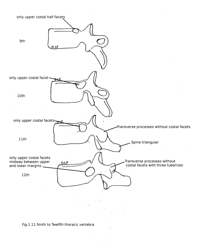
It shows all features of atypical thoracic vertebra but some exceptions are visible. Body shows only upper costal half facets for head of ninth ribs. Lower costal facets are absent.
Tenth thoracic vertebra
It shows only upper costal facets on body (near upper margin) which articulate with heads of tenth ribs.
Eleventh thoracic vertebra
It shows only upper costal facets on body (slightly low position near upper margin) which articulate with heads of eleventh ribs. Transverse processes are small and without any costal facets. Spine is triangular in shape.
Twelfth thoracic vertebra
Its body, pedicles, transverse processes and spine resembles lumbar vertebra. It shows only upper costal facets on body (midway between upper and lower margins) which articulate with heads of twelfth ribs. Transverse processes are small and without any costal facets having superior, inferior and lateral tubercles. Inferior articular facets are convex facing antero-laterally. Spine is triangular in shape.
