ARCHES OF FOOT
Functions of foot are 1) support body weight during standing 2) acts as a lever during running and walking.
Foot is developed in such a way so it can perform all above functions of walking and running. Bones of foot are formed by small bones containing multiple joints. So foot becomes pliable to adjust on uneven surface. Elastic arches are present in foot to sustain body weight and stress during movements.
Two types of arches are present in foot 1) longitudinal 2) transverse
Functions of arches :
1) It causes distribution of body weight in foot. Weight transmitted to foot half to calcaneus and half part to heads of five metatarsals. Then anterior part divides weight into six equal parts two for great toe and one each for second to fifth metatarsal heads.
2) Segmented lever formed by small bones helps in propulsive action.
3) Concave plantar aspect protect vessels and nerves of foot.
4) Arches make foot pliable on uneven ground
1) LONGITUDINAL ARCH
Two longitudinal arches present in foot a) Medial longitudinal arch and b) Lateral longitudinal arch.
a) Medial longitudinal arch
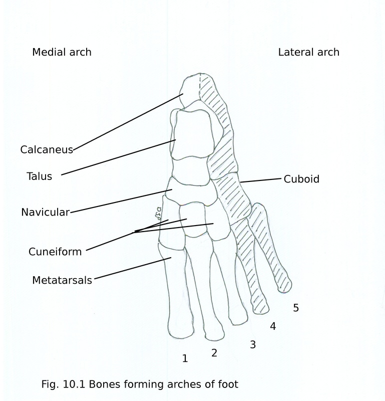
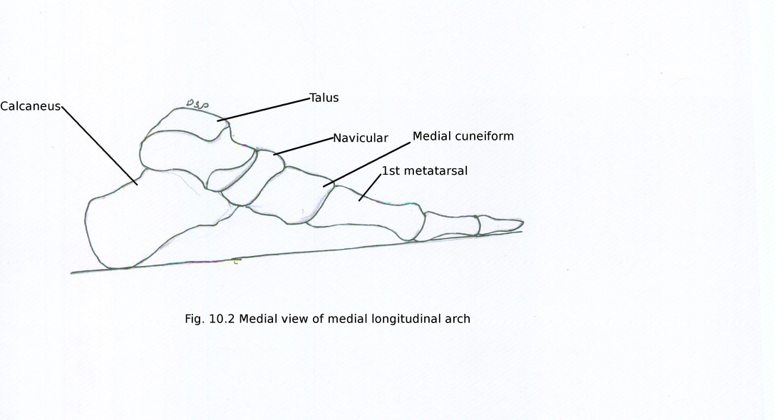
Bones forming arch : Bones of the arch are calcaneus, head of talus, navicular, three cuneiform, medial three metatarsals.
Summit : It is formed by talus.
Pillars : Posterior pillar is formed by medial tubercle of calcaneus and anterior pillar is formed by heads of medial three metatarsals.
Joints : Joints are talocalcaneonavicular and subtalar.
Factors maintaining medial longitudinal arch :
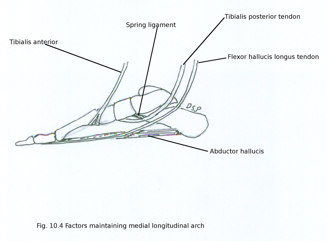
Bones : Shape of bone play a main role in maintenance of arches. Head of talus articulating with navicular bone supported below by sustentaculum tali of calcaneus.
Ligaments : Plantar ligaments bind bones of arches. Important ligament in it is plantar calcaneonavicular or spring ligament support head of talus. Other ligaments are interosseous ligaments connecting adjacent bones, anterior fibres of deltoid ligament from tibia to navicular and interosseous talocalcanean ligament. These act as intersegmental ties.
Muscles, tendons and aponeurosis : Flexor hallucis longus work as a bowstring. Other muscles are flexor digitorum longus, abductor hallucis and medial half of flexor digitorum brevis. Tibialis posterior help in inversion of foot and support from below. Plantar aponeurosis is main binding structure. Tibialis anterior along with deltoid ligament support arch from above
Two longitudinal arches present in foot a) Medial longitudinal arch and b) Lateral longitudinal arch.
a) Medial longitudinal arch
Bones forming arch : Bones of the arch are calcaneus, head of talus, navicular, three cuneiform, medial three metatarsals.
Summit : It is formed by talus.
Pillars : Posterior pillar is formed by medial tubercle of calcaneus and anterior pillar is formed by heads of medial three metatarsals.
Joints : Joints are talocalcaneonavicular and subtalar.
Factors maintaining medial longitudinal arch :
Bones : Shape of bone play a main role in maintenance of arches. Head of talus articulating with navicular bone supported below by sustentaculum tali of calcaneus.
Ligaments : Plantar ligaments bind bones of arches. Important ligament in it is plantar calcaneonavicular or spring ligament support head of talus. Other ligaments are interosseous ligaments connecting adjacent bones, anterior fibres of deltoid ligament from tibia to navicular and interosseous talocalcanean ligament. These act as intersegmental ties.
Muscles, tendons and aponeurosis : Flexor hallucis longus work as a bowstring. Other muscles are flexor digitorum longus, abductor hallucis and medial half of flexor digitorum brevis. Tibialis posterior help in inversion of foot and support from below. Plantar aponeurosis is main binding structure. Tibialis anterior along with deltoid ligament support arch from above
b) Lateral longitudinal arch
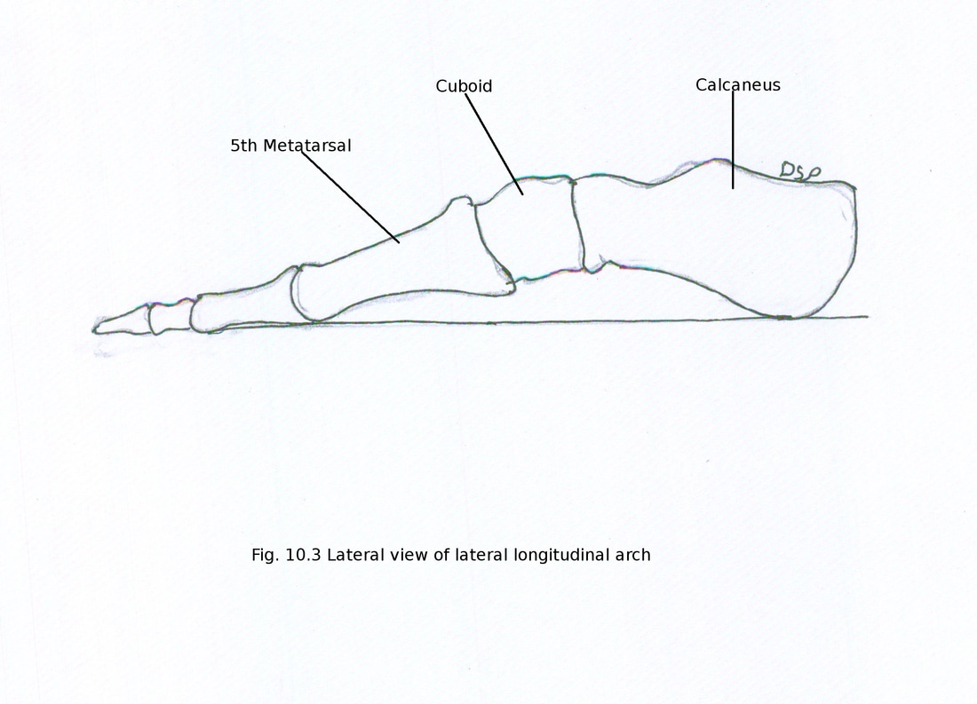
Bones forming arch : Bones of the arch are calcaneus, cuboid, fourth and fifth metatarsals.
Summit : It is formed by subtalar joint.
Pillars : Posterior pillar is formed by medial tubercle of calcaneus and anterior pillar is formed by heads of fourth and fifth metatarsals.
Joints : Main joint is calcaneocuboid.
Factors maintaining lateral longitudinal arch :
Bones : Shape of bone play a main role in maintenance of arches. Triangular projection of cuboid from its inferior margin of proximal articular surface articulate with anterior articular surface of calcaneus from below.
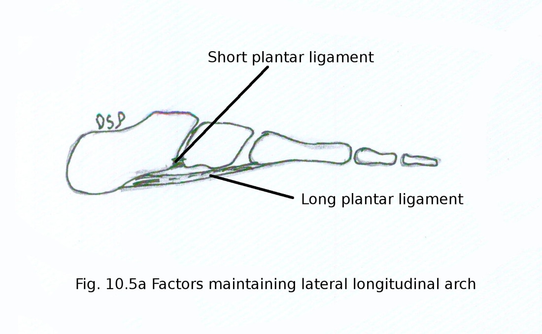
Ligaments : Plantar ligaments bind bones of arches. Lateral part of plantar aponeurosis main binding agent. Long plantar ligament support joints between calcaneus, cuboid and metatarsals and short plantar ligaments supports calcaneocuboid joint from below. These act as intersegmental ties.
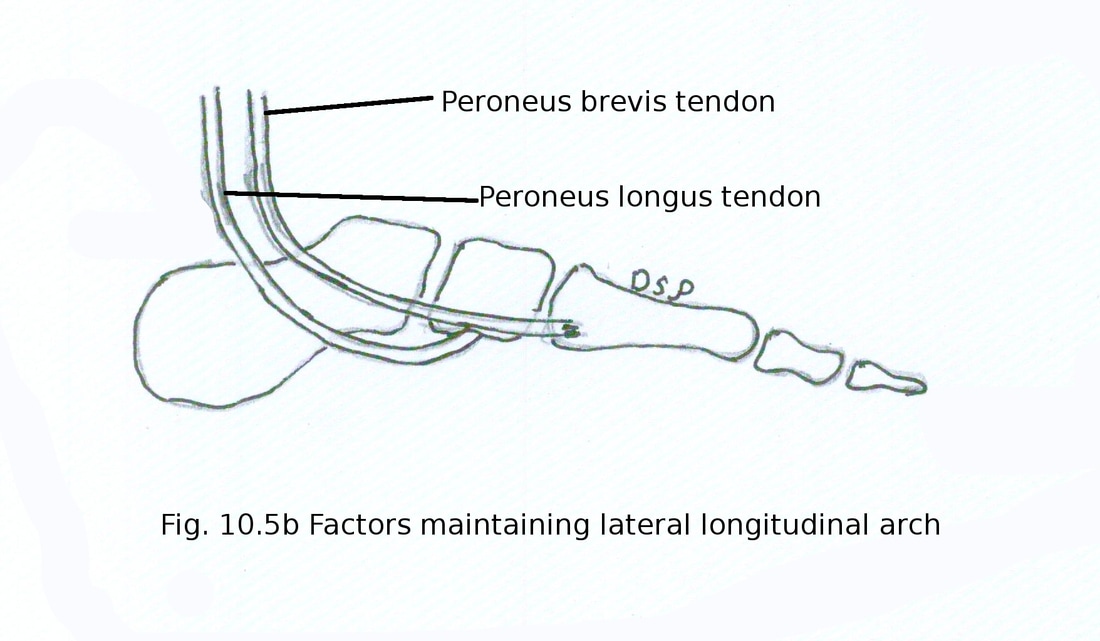
Muscles, tendons and aponeurosis : Lateral part of flexor digitorum brevis, tibialis posterior, flexor digiti minimi brevis, abductor digiti minimi act as tie beams. Tendons of peroneus brevis and peroneus tertius supports from above. Peroneus longus tendon maintains lateral arch by supporting arch from below. Plantar aponeurosis acts as tie beams
Bones forming arch : Bones of the arch are calcaneus, cuboid, fourth and fifth metatarsals.
Summit : It is formed by subtalar joint.
Pillars : Posterior pillar is formed by medial tubercle of calcaneus and anterior pillar is formed by heads of fourth and fifth metatarsals.
Joints : Main joint is calcaneocuboid.
Factors maintaining lateral longitudinal arch :
Bones : Shape of bone play a main role in maintenance of arches. Triangular projection of cuboid from its inferior margin of proximal articular surface articulate with anterior articular surface of calcaneus from below.
Ligaments : Plantar ligaments bind bones of arches. Lateral part of plantar aponeurosis main binding agent. Long plantar ligament support joints between calcaneus, cuboid and metatarsals and short plantar ligaments supports calcaneocuboid joint from below. These act as intersegmental ties.
Muscles, tendons and aponeurosis : Lateral part of flexor digitorum brevis, tibialis posterior, flexor digiti minimi brevis, abductor digiti minimi act as tie beams. Tendons of peroneus brevis and peroneus tertius supports from above. Peroneus longus tendon maintains lateral arch by supporting arch from below. Plantar aponeurosis acts as tie beams
2) TRANSVERSE ARCH
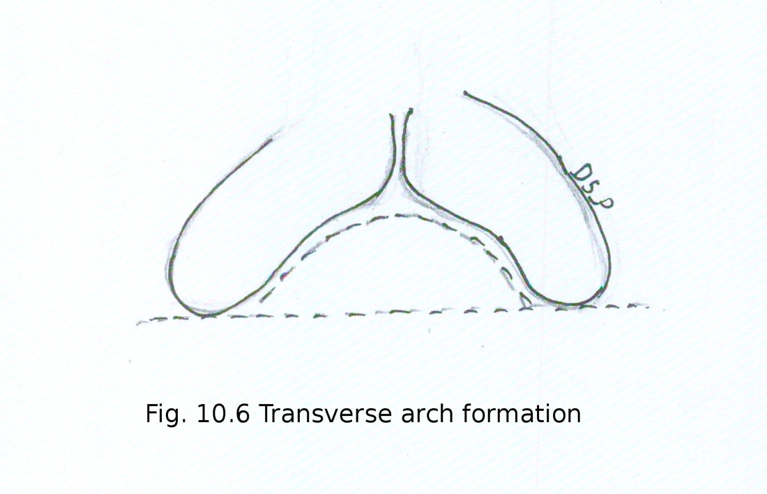
Bones forming arch : Anterior transverse arch is formed by bases of five metatarsal bones and posterior transverse arch is formed by tarsals, metatarsals like cuboid and cuneiform. Posterior arch is incomplete arch in each foot forming half dome and is complete when two foot are brought together.
Factors maintaining transverse arch :
Bones : Shape of bone play a main role in maintenance of arches. Wedge shaped three cuneiform bones with larger dorsal and smaller plantar surface and bases of middle three metatarsal bones are bony factors.
Ligaments : Plantar ligaments bind bones of arches. Ligament are deep transverse ligaments between plantar surface of tarsal and metatarsal bones, intrinsic plantar ligaments act as intersegmental ties.
Muscles, tendons and aponeurosis : Dorsal interossei, oblique and transverse heads of adductor hallucis acts as intersegmental ties. Peroneus longus and tibialis posterior tendon maintains transverse arch from below and acts as tie beams. Peroneus tertius, peroneus brevis and tibialis anterior on medial side supports as a sling.
Applied anatomy of arches of foot :
1. Pes planus :Absence of arches causes pes planus (flat foot). Flat foot causes compression of nerves, vessels. Pain in foot because of compression of nerves. Clumsy gait developed with loss of spring action of foot this leads to trauma and osteoarthritis.
2. Claw foot or pes cavus : Exaggeration of longitudinal arches and foot remain in plantar flexed condition. Also there is dorsiflexion at metatarsophalangeal joints and plantar flexion at interphalangeal joints. It occurs in spina bifida and poliomyelitis.
3. Club foot : Following types of club foot are seen.
a) Talipes equines : Person walks on toes and heel raised.
b) Talipes calcaneus : Person walks on heel with toes raised.
c) Talipes varus : Person walks on outer border of foot. Foot remains in inverted and adducted position.
d) Talipes valgus : Person walks on inner border of foot. Foot remains in inverted and abducted position.
Talipes equinovarus in which person walks on outer margin anterior part of foot. Foot remains inverted, adducted and plantar flexed.
