Back of Thigh And Popliteal fossa
It extends from lower part of gluteal region to back of knee joint up to popliteal fossa. It is also a flexor compartment of thigh. It lies in between laterally lateral intermuscular septum and medially posterior intermuscular compartment which is incomplete. In the floor adductor magnus and vastus lateralis lies. Fascia lata covers back of thigh.
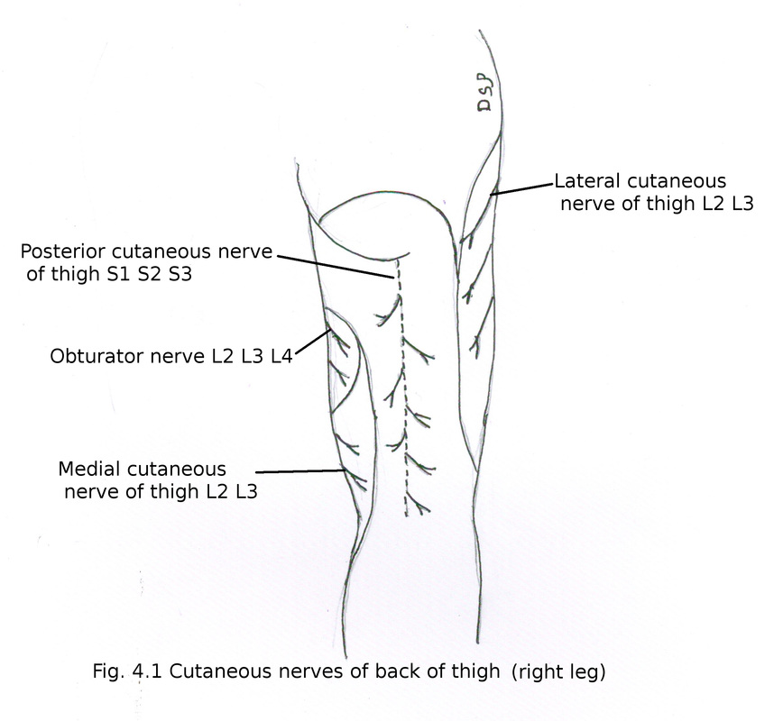
Cutaneous nerves
Skin of back of thigh supplied by following cutaneous nerves
1) Posterior femoral cutaneous nerve
Formed by dorsal branches of S1,S2 and ventral branches of S2, S3. It passes through greater sciatic foramen below piriformis medial to sciatic nerve. It goes down to back of thigh deep to fascia latae. In relation with roof of popliteal fossa accompanies small saphenous vein and communicate with sural nerve.
Branches 1) Gluteal : Supply skin of lower and lateral part of gluteal region. 2) Perineal : Supply superomedial skin of thigh and posterior part of scrotum or labium majus. 3) Perforating : Supply skin of back of thigh, popliteal fossa and upper part of back of leg.
2) Cutaneous branches of obturator nerve
Supply skin of back of thigh in upper part.
3) Medial branches of anterior cutaneous nerve of thigh
Supply skin of medial part back of thigh in lower part.
4) Branch of Obturator nerve (L2, L3, L4)
Anterior division of obturator nerve supply medial and lower part of thigh.
5) Lateral cutaneous nerve of thigh
Supply skin of medial part of upper aspect of thigh.
It extends from lower part of gluteal region to back of knee joint up to popliteal fossa. It is also a flexor compartment of thigh. It lies in between laterally lateral intermuscular septum and medially posterior intermuscular compartment which is incomplete. In the floor adductor magnus and vastus lateralis lies. Fascia lata covers back of thigh.
Cutaneous nerves
Skin of back of thigh supplied by following cutaneous nerves
1) Posterior femoral cutaneous nerve
Formed by dorsal branches of S1,S2 and ventral branches of S2, S3. It passes through greater sciatic foramen below piriformis medial to sciatic nerve. It goes down to back of thigh deep to fascia latae. In relation with roof of popliteal fossa accompanies small saphenous vein and communicate with sural nerve.
Branches 1) Gluteal : Supply skin of lower and lateral part of gluteal region. 2) Perineal : Supply superomedial skin of thigh and posterior part of scrotum or labium majus. 3) Perforating : Supply skin of back of thigh, popliteal fossa and upper part of back of leg.
2) Cutaneous branches of obturator nerve
Supply skin of back of thigh in upper part.
3) Medial branches of anterior cutaneous nerve of thigh
Supply skin of medial part back of thigh in lower part.
4) Branch of Obturator nerve (L2, L3, L4)
Anterior division of obturator nerve supply medial and lower part of thigh.
5) Lateral cutaneous nerve of thigh
Supply skin of medial part of upper aspect of thigh.
Contents
Muscles : Hamstring muscles, short head of biceps femoris
Nerves : Sciatic nerve, Posterior femoral cutaneous nerve
Arteries : Anastomosis on back of thigh
Muscles
Hamstring muscles are semitendinosus, semimembranosus, long head of biceps femoris and ischial fibres of adductor magnus.
Characteristic features shown by hamstring muscles are :
a) They show origin from ischial tuberosity.
b) They show insertion on tibia or fibula.
c) They are supplied by tibial division of sciatic nerve.
d) They help in flexion of knee joint and extension of hip joint.
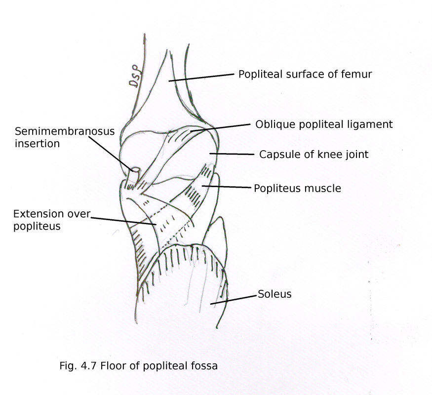
Semimembranosus
Origin : It shows origin from upper and lateral part of quadrilateral area of ischial tuberosity. It is membranous in upper part and fleshy in lower part.
Insertion : It shows insertion on medial condyle of tibia on its posterior aspect in a horizontal groove.
It shows three expansions 1) a fibrous band named oblique popliteal ligament which goes upward and laterally posterior to capsule of knee joint shows attachment on inter-condylar line and lateral condyle of femur 2) fibrous expansion downward and laterally shows attachment on soleal line of tibia 3) few fibres goes downward shows attachment on medial border of upper part of tibia.
Nerve supply : It receives nerve supply from tibial part of sciatic nerve L5, S1, S2.
Muscles : Hamstring muscles, short head of biceps femoris
Nerves : Sciatic nerve, Posterior femoral cutaneous nerve
Arteries : Anastomosis on back of thigh
Muscles
Hamstring muscles are semitendinosus, semimembranosus, long head of biceps femoris and ischial fibres of adductor magnus.
Characteristic features shown by hamstring muscles are :
a) They show origin from ischial tuberosity.
b) They show insertion on tibia or fibula.
c) They are supplied by tibial division of sciatic nerve.
d) They help in flexion of knee joint and extension of hip joint.
Semimembranosus
Origin : It shows origin from upper and lateral part of quadrilateral area of ischial tuberosity. It is membranous in upper part and fleshy in lower part.
Insertion : It shows insertion on medial condyle of tibia on its posterior aspect in a horizontal groove.
It shows three expansions 1) a fibrous band named oblique popliteal ligament which goes upward and laterally posterior to capsule of knee joint shows attachment on inter-condylar line and lateral condyle of femur 2) fibrous expansion downward and laterally shows attachment on soleal line of tibia 3) few fibres goes downward shows attachment on medial border of upper part of tibia.
Nerve supply : It receives nerve supply from tibial part of sciatic nerve L5, S1, S2.
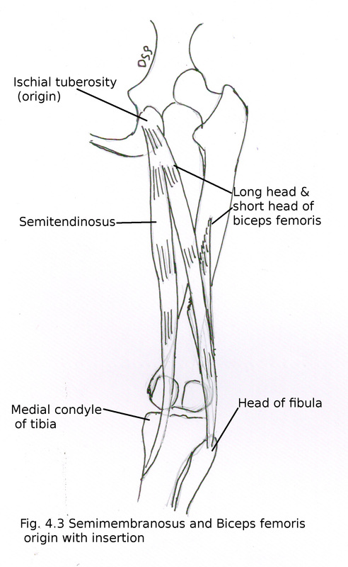
Semitendinosus
Origin : It shows origin along with long head of biceps femoris from lower and medial part of quadrilateral area of ischial tuberosity. It is fleshy in upper part and forms a tendon in lower part.
Insertion : It shows insertion of its tendon on upper part of medial surface of tibia posterior to insertion of sartorius and gracilis.
Nerve supply : It receives nerve supply from tibial part of sciatic nerve (L5, S1, S2).
Biceps femoris
Origin : It shows two heads of origin long and short. Long head is part of hamstring muscle. Long head shows origin along with semitendinosus from lower and medial part of quadrilateral area of ischial tuberosity. It goes downward and laterally to join with its short head. Short head shows origin from lateral lip of linea aspera from its lower part and upper 2/3 part of lateral supracondylar line.
Insertion : It shows insertion of its tendon on head of fibula in front of styloid process.
Nerve supply : Long head receives nerve supply from tibial part of sciatic nerve (L5, S1, S2), short head receives nerve supply from common peroneal part of sciatic nerve (L5, S1, S2).
Action of Hamstring muscles
Flexion at knee joint. During standing and walking help in extension of hip joint. It also help in medial rotation of tibia on femur by semimembranosus and semitendinosus while lateral rotation of tibia by biceps femoris.
Nerves
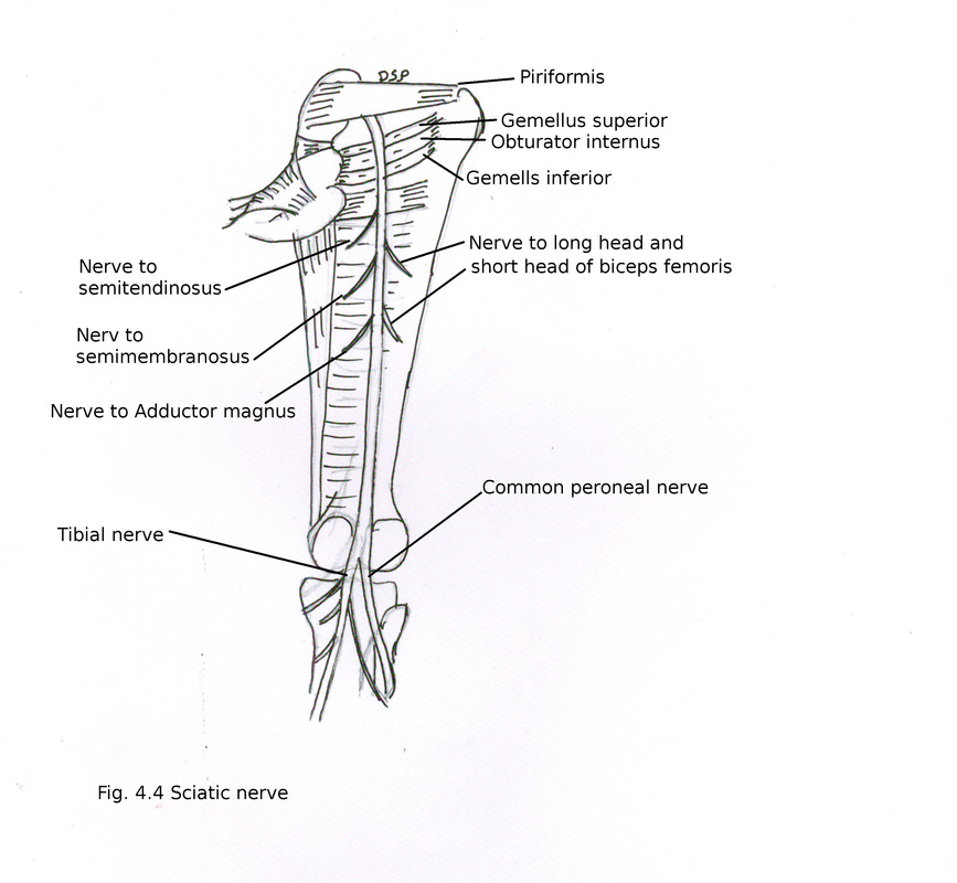
Sciatic nerve
It is thickest nerve. Sacral plexus forms sciatic nerve. It shows two parts tibial and common peroneal. Tibial part formed by ventral division of anterior primary rami of L4,L5,S1,S2,S3. Common peroneal part formed by dorsal division of anterior primary rami of L4,L5,S1,S2. It passes straight downward from lower margin of piriformis upto upper angle of popliteal fossa deep to long head of biceps femoris and over the surface of adductor magnus. It is accompanied medially by posterior femoral cutaneous nerve and inferior gluteal artery. Then it divides into two terminal branches 1) Tibial and 2) Common peroneal
Branches in back of thigh : 1) Branches to hamstring muscles arises from medial side of nerve from its tibial component 2) Branch to short head of biceps femoris from its lateral side from its common peroneal component 3) Articular branches to hip joint.
Flexion at knee joint. During standing and walking help in extension of hip joint. It also help in medial rotation of tibia on femur by semimembranosus and semitendinosus while lateral rotation of tibia by biceps femoris.
Nerves
Sciatic nerve
It is thickest nerve. Sacral plexus forms sciatic nerve. It shows two parts tibial and common peroneal. Tibial part formed by ventral division of anterior primary rami of L4,L5,S1,S2,S3. Common peroneal part formed by dorsal division of anterior primary rami of L4,L5,S1,S2. It passes straight downward from lower margin of piriformis upto upper angle of popliteal fossa deep to long head of biceps femoris and over the surface of adductor magnus. It is accompanied medially by posterior femoral cutaneous nerve and inferior gluteal artery. Then it divides into two terminal branches 1) Tibial and 2) Common peroneal
Branches in back of thigh : 1) Branches to hamstring muscles arises from medial side of nerve from its tibial component 2) Branch to short head of biceps femoris from its lateral side from its common peroneal component 3) Articular branches to hip joint.
Posterior femoral cutaneous nerve
It is formed by dorsal branches of sacral S1, S2 and ventral branches of sacral S2, S3. It goes out of pelvis through greater sciatic foramen. Goes just below lower border of piriformis vertically downward medial to sciatic nerve. It lies superficial to long head of biceps femoris in back of thigh. In relation with roof of popliteal fossa it pierces popliteal fascia. Accompanies small saphenous vein and communicate with sural nerve.
Branches 1) Gluteal : Supply skin of lower and lateral part of gluteal region. 2) Perineal : Supply superomedial skin of thigh and posterior part of scrotum or labium majus. 3) Perforating : Supply skin of back of thigh, popliteal fossa and upper part of back of leg.
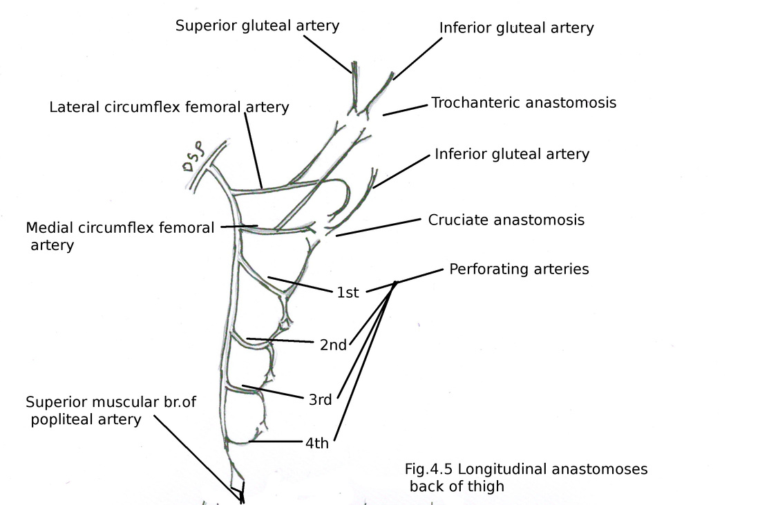
Anastomosis on back of thigh
Longitudinal anastomosis : It is formed by perforating branches of profunda femoris artery. Perforating branches of profunda femoris pierce adductor magnus muscle. Then gives ascending and descending branches. Four perforating branches anastomose with each other by its ascending and descending branches. Ascending branch of 1st perforating artery joins with cruciate anastomosis. Descending branch of 4th perforating artery joins with superior muscular branch of popliteal artery.
It is formed by dorsal branches of sacral S1, S2 and ventral branches of sacral S2, S3. It goes out of pelvis through greater sciatic foramen. Goes just below lower border of piriformis vertically downward medial to sciatic nerve. It lies superficial to long head of biceps femoris in back of thigh. In relation with roof of popliteal fossa it pierces popliteal fascia. Accompanies small saphenous vein and communicate with sural nerve.
Branches 1) Gluteal : Supply skin of lower and lateral part of gluteal region. 2) Perineal : Supply superomedial skin of thigh and posterior part of scrotum or labium majus. 3) Perforating : Supply skin of back of thigh, popliteal fossa and upper part of back of leg.
Anastomosis on back of thigh
Longitudinal anastomosis : It is formed by perforating branches of profunda femoris artery. Perforating branches of profunda femoris pierce adductor magnus muscle. Then gives ascending and descending branches. Four perforating branches anastomose with each other by its ascending and descending branches. Ascending branch of 1st perforating artery joins with cruciate anastomosis. Descending branch of 4th perforating artery joins with superior muscular branch of popliteal artery.
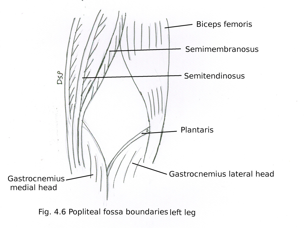
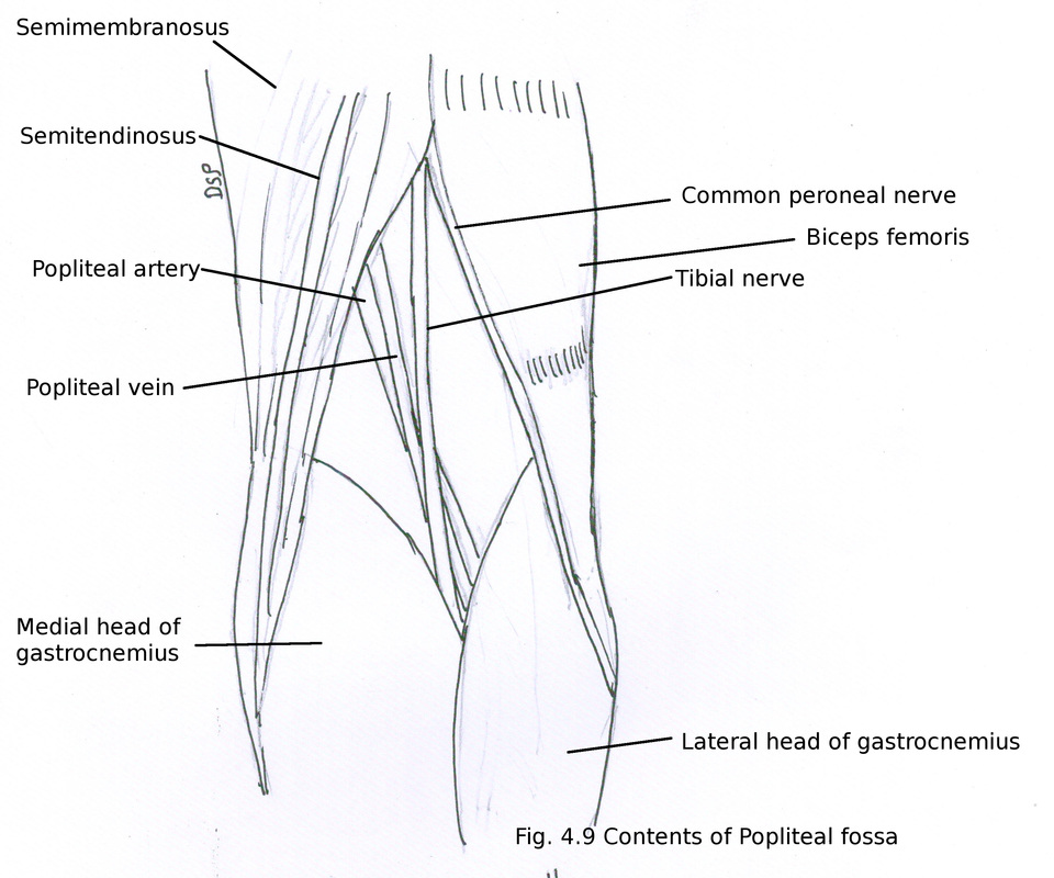
Popliteal fossa
It is a diamond shaped space present on back of knee joint.
Boundaries :
Superomedially : Semitendinosus and semimembranosus
Superolaterally : Tendon of biceps femoris muscle
Inferomedially : Medial head of gastrocnemius
Inferolaterally : Lateral head of gastrocnemius and plantaris
It is a diamond shaped space present on back of knee joint.
Boundaries :
Superomedially : Semitendinosus and semimembranosus
Superolaterally : Tendon of biceps femoris muscle
Inferomedially : Medial head of gastrocnemius
Inferolaterally : Lateral head of gastrocnemius and plantaris
Floor (anterior wall) : It is formed from above downward by Popliteal surface of femur, capsule of knee joint and oblique popliteal ligament, posterior surface of upper end of tibia, fascia covering popliteus.
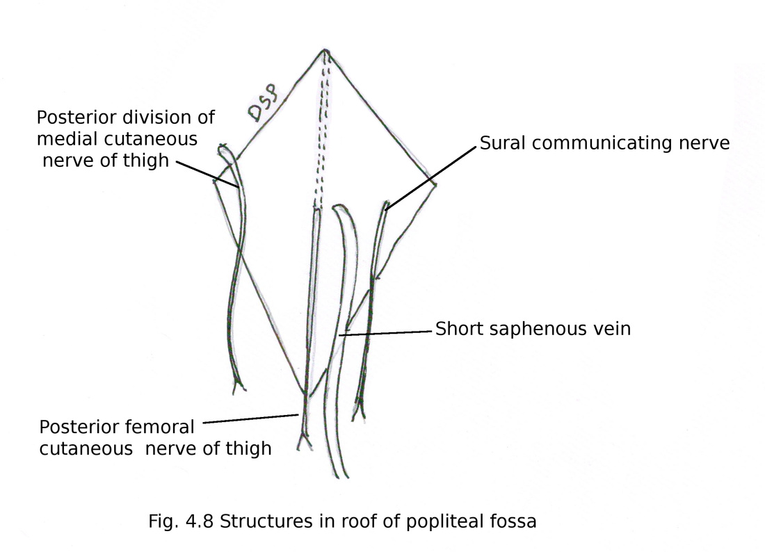
Roof (anterior wall) : Popliteal fascia
Roof also contains short saphenous vein, cutaneous nerves terminal part of posterior femoral cutaneous nerve, posterior division of medial cutaneous nerve of thigh, sural communicating nerve. All these structures pierce roof except posterior division of medial cutaneous nerve of thigh.
Contents :
1) Popliteal artery
2) Popliteal vein
3) Tibial nerve
4) Common peroneal nerve
5) Popliteal lymphnodes
6) Popliteal fat
other small structures
7) Terminal part of small saphenous vein
8) Terminal part of Posterior femoral cutaneous nerve
9) Descending genicular branch of posterior division of obturator nerve
1) Popliteal artery
It is continuation of femoral artery after its exit from 5th opening in adductor magnus muscle upto lower margin of popliteus muscle where it divides into anterior and posterior tibial arteries. Course is obliquely downward and laterally. Popliteal vein runs alongwith artery. Vein crosses artery from lateral to medial superficial to artery. So vein lies posterolateral to artery in upper part and posteromedial to artery in lower part. Tibial nerve goes vertically downward from upper angle of popliteal fossa to lower angle of popliteal fossa superficial to vein.
Branches : (except terminal)
1) Muscular branches supply muscles adductor magnus, hamstring.
2) Articular branches supply knee joint by genicular arteries a) superior medial b) superior lateral near lower femoral condyle and c) inferior medial d) inferior lateral near upper end of tibial condyles and e) middle genicular artery pierce oblique popliteal ligament supply cruciate ligament and synovial membrane.
3) Cutaneous branches after piercing roof of fossa supply back of leg and sural branch runs alongwith small saphenous vein.
1) Popliteal artery
2) Popliteal vein
3) Tibial nerve
4) Common peroneal nerve
5) Popliteal lymphnodes
6) Popliteal fat
other small structures
7) Terminal part of small saphenous vein
8) Terminal part of Posterior femoral cutaneous nerve
9) Descending genicular branch of posterior division of obturator nerve
1) Popliteal artery
It is continuation of femoral artery after its exit from 5th opening in adductor magnus muscle upto lower margin of popliteus muscle where it divides into anterior and posterior tibial arteries. Course is obliquely downward and laterally. Popliteal vein runs alongwith artery. Vein crosses artery from lateral to medial superficial to artery. So vein lies posterolateral to artery in upper part and posteromedial to artery in lower part. Tibial nerve goes vertically downward from upper angle of popliteal fossa to lower angle of popliteal fossa superficial to vein.
Branches : (except terminal)
1) Muscular branches supply muscles adductor magnus, hamstring.
2) Articular branches supply knee joint by genicular arteries a) superior medial b) superior lateral near lower femoral condyle and c) inferior medial d) inferior lateral near upper end of tibial condyles and e) middle genicular artery pierce oblique popliteal ligament supply cruciate ligament and synovial membrane.
3) Cutaneous branches after piercing roof of fossa supply back of leg and sural branch runs alongwith small saphenous vein.
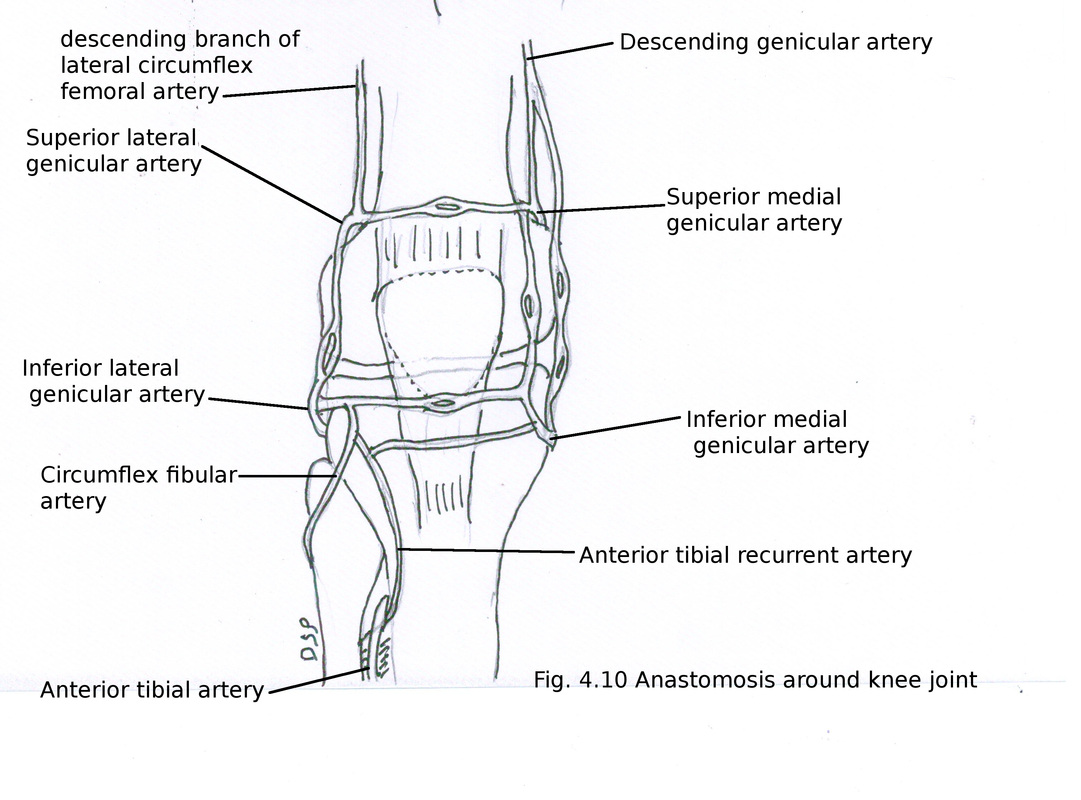
Genicular anastomosis:
It is an anastomosis around patella and tibial and lower femoral condyles. It supply capsule of knee, synovial membrane, bone. It is formed by superior medial genicular, superior lateral genicular, inferior medial genicular, inferior lateral genicular, middle genicular arteries.
Superior genicular arteries are branches of popliteal artery. It is near femoral condyles. Superior medial genicular artery arches over medial head of gastrocnemius and deep to tendon of adductor magnus and anastomoses with descending genicular branch of femoral artery, inferior medial genicular artery and superior lateral genicular artery. Lateral superior genicular artery passes deep to tendon of biceps femoris across lateral head of gastrocnemius gives out superficial and deep branch. Superficial branch anastomoses with descending branch of lateral circumflex femoral artery and inferior lateral genicular artery. Deep branch anastomoses with medial superior genicular artery.
Inferior genicular arteries are medial and lateral genicular arteries. These are branches of popliteal artery. These lies deep to gastrocnemius. Medial branch goes below medial tibial condyle deep to tibial collateral ligament. Medial branch anastomoses with superior medial genicular artery, saphenous branch of descending genicular artery, lateral inferior genicular artery and anterior tibial recurrent artery. Lateral branch lies along lateral tibial condyle near head of fibula covered by fibular collateral ligament. Lateral branch anastomoses with superior lateral genicular artery, anterior and posterior tibial recurrent branches of anterior tibial artery and circumflex fibular branch of posterior tibial artery, inferior medial genicular artery.
Applied anatomy : popliteal aneurysm present in mid-line of popliteal fossa as pulsatile swelling.
2) Popliteal vein
It is formed near lower margin of popliteus. Formed by joining veins which accompanies anterior and posterior tibial arteries. It goes up after crossing popliteal artery from medial to lateral side and superficial to artery (posteriorly) in popliteal fossa. Popliteal vein goes up as femoral vein after passing through opening in adductor magnus muscle. Tributaries of it are small saphenous vein, small muscular veins and tributaries which which runs with branches of popliteal artery.
3) Tibial nerve
It is a branch of sciatic nerve. Formed by ventral branches of L4, L5, S1, S2, S3. It goes vertically downward from superior angle to inferior angle of popliteal fossa. It crosses popliteal artery from lateral to medial side and lies superficial to popliteal artery in popliteal fossa. Popliteal vein lies in between artery and nerve. In lower part of popliteal fossa covered by two heads of gastrocnemius. Here it enters in back of leg alongwith posterior tibial vessels.
Branches:
1) Muscular : It supply gastrocnemius, soleus, plantaris, popliteus. Muscular branches arise from lateral aspect of tibial nerve except branch to medial head of gastrocnemius. Branch to popliteus also supply superior tibio-fibular joint, inferior tibio-fibular joint, interosseous membrane and tibialis posterior.
2) Articular : It mainly supply knee joint by three branches superior medial genicular, inferior medial genicular nerve and medial genicular nerve. All these branches runs alongwith arteries of same name. These joins with branch of obturator nerve to supply oblique popliteal ligament.
3) Cutaneous : Sural nerve L5, S1, S2 joins with sural communicating nerve branch of common peroneal nerve. Supply posterior and lateral aspect of leg in lower one third part.
4) Vascular branch : Sympathetic nerve from T10, T11, T12, L1, L2 post ganglionic branches vasomotor fibres to popliteal vessels.
4) Common peroneal nerve
It is branch of sciatic nerve. Formed by dorsal branches of L4, L5 and ventral rami of S1, S2. It goes vertically downward laterally from deep surface of long head of biceps. It crosses plantaris, lateral head of gastrocnemius superficially and crosses neck of fibula. Then it goes anteriorly after crossing neck of fibula. Gives out two terminal branches superficial peroneal nerve and deep peroneal nerve.
Branches in popliteal fossa (other than terminal branches) :
1) Articular : It mainly supply knee joint by three branches superior lateral genicular, inferior lateral genicular nerve and recurrent genicular nerve. All these branches runs alongwith arteries of same name. Recurrent genicular nerve supply anterolateral part of knee joint and superior tibio-fibular joint.
2) Cutaneous : Sural communicating nerve and lateral sural nerve (lateral cutaneous nerve of calf) are cutaneous branches of common peroneal nerve. Supply upper anterior, posterior and lateral aspect of leg.
Applied anatomy : Fracture neck of fibula causes injury to common peroneal nerve. Effects of injury are paralysis of peroneal and extensor muscles of leg causing foot drop. Position of foot inverted, plantar flexed and adducted. Sensory loss over dorsum of foot and lower one third of front of leg.
5) Popliteal lymphnodes
These are six in numbers. One lies near termination of small saphenous vein draining lymph from area of small saphenous vein. One on posterior aspect of knee joint and popliteal artery draining lymph from knee joint and area of genicular vessels.. Other lymphnode lies in relation with popliteal vessels draining lymph from area of anterior and posterior tibial vessels.
Lymph vessels goes to deep inguinal lymphnodes alongwith femoral vessels.
6) Popliteal fat
Popliteal fat present in between vessels and nerves and other contents of fossa.
7) Terminal part of small saphenous vein
Small saphenous vein after piercing roof of popliteal fossa drains into popliteal vein.
8) Terminal part of Posterior femoral cutaneous nerve
In relation with roof of popliteal fossa accompanies small saphenous vein and communicate with sural nerve. After piercing roof of popliteal fossa supply skin of popliteal fossa and upper part of back of leg.
9) Descending genicular branch of posterior division of obturator nerve
It is a branch of posterior division of obturator nerve. It goes alongwith popliteal artery and after piercing oblique popliteal ligament supply capsule of knee joint.
