AXILLA
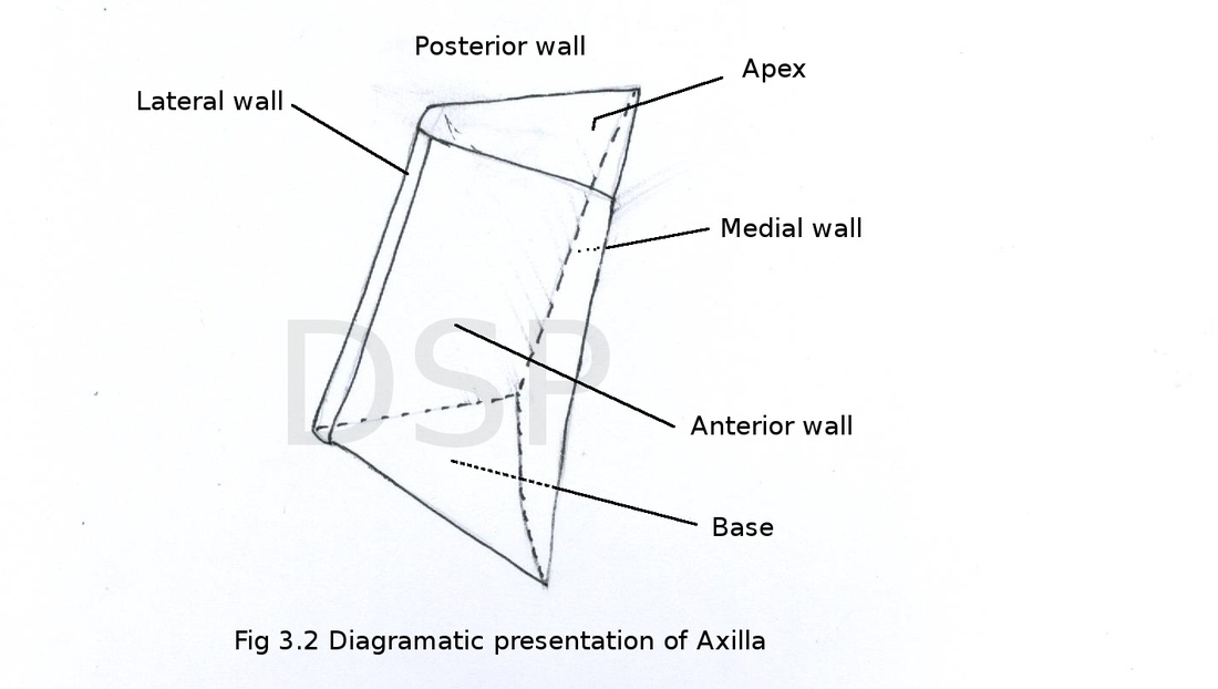
Axilla is a pyramid shaped space between medial surface of upper arm and lateral surface of thoracic wall. It has apex, base, anterior wall, posterior wall, medial wall and lateral wall.
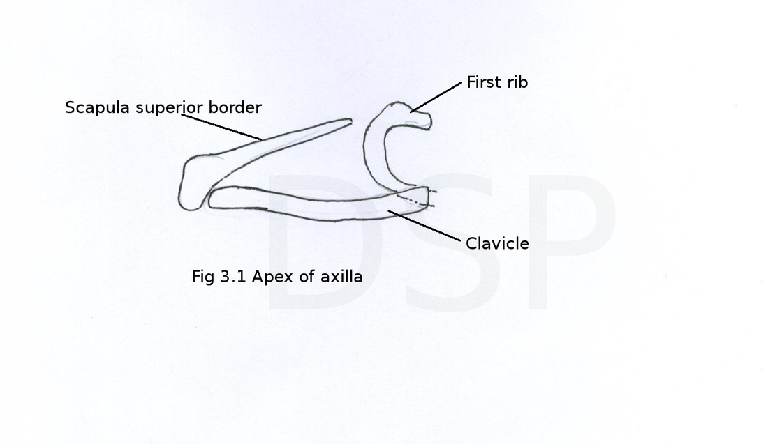
Apex: It is a triangular shaped space bounded by three bones posteriorly by superior border of scapula anteriorly by clavicle and medialy by outer margin of a 1st rib. Cords of brachial plexus, axillary vessels, some lymphatic vessels and long thoracic nerve passes through this space.
Base: It is concave shaped bounded by anterior and posterior axillary folds below by skin and fascia.
Anterior wall: It is formed by pectoralis major, pectoralis minor, subclavius and clavipectoral fascia.
Posterior wall : Formed by subscapularis, lattissimus dorsi and teres major muscles.
Medial wall: Formed by upper 4 ribs with interostal spaces and intercostal muscles also by upper part of serratus anterior muscle.
Lateral wall: Formed by convergence of anterior and posterior wall on intertubercular sulcus of humerus along with long tendon of biceps brachii and coracobrachialis with short head of biceps brachii.
Contents of axilla
Axillary artery and its branches , Axillary vein and its tributaries, Brachial plexus cords, branches from brachial plexus, Long thoracic nerve , Intercosto brachial nerve, Axillary fat and axillary group of lymph nodes.
Axilla is a pyramid shaped space between medial surface of upper arm and lateral surface of thoracic wall. It has apex, base, anterior wall, posterior wall, medial wall and lateral wall.
Apex: It is a triangular shaped space bounded by three bones posteriorly by superior border of scapula anteriorly by clavicle and medialy by outer margin of a 1st rib. Cords of brachial plexus, axillary vessels, some lymphatic vessels and long thoracic nerve passes through this space.
Base: It is concave shaped bounded by anterior and posterior axillary folds below by skin and fascia.
Anterior wall: It is formed by pectoralis major, pectoralis minor, subclavius and clavipectoral fascia.
Posterior wall : Formed by subscapularis, lattissimus dorsi and teres major muscles.
Medial wall: Formed by upper 4 ribs with interostal spaces and intercostal muscles also by upper part of serratus anterior muscle.
Lateral wall: Formed by convergence of anterior and posterior wall on intertubercular sulcus of humerus along with long tendon of biceps brachii and coracobrachialis with short head of biceps brachii.
Contents of axilla
Axillary artery and its branches , Axillary vein and its tributaries, Brachial plexus cords, branches from brachial plexus, Long thoracic nerve , Intercosto brachial nerve, Axillary fat and axillary group of lymph nodes.
Axillary artery
It is a continuation of third part of subclavian artery. It extends from outer margin of first rib to lower margin of teres major muscle afterwards it continue as brachial artery this artery is divided into 3 parts by pectoralis minor muscle. First part proximal to muscle, second part posterior to muscle and third part distal to the muscle.
Relations of axillary artery
First part
Anterior relation: skin, superficial fascia, platysma and supraclavicular nerves deep fascia pectoralis major muscle, clavipectoral fascia, a communicating loop between lateral and medial pectoral nerve. Axillary artery
Posterior relations: first two digitations of serratus anterior muscle with its nerve, medial cord of brachial plexus and it's medial pectoral branch.
Lateral relations: lateral and posterior cords of brachial plexus.
Medial relations : axillary vein.
Second part
Anterior relations: skin, fascia, pectoralis major, pectoralis minor.
Posterior relations: Subscapularis muscle, posterior cord of brachial plexus.
Lateral relations: lateral cord
Medial relations: medial cord, axillary vein , medial pectoral nerve.
Third part
Anterior relations: skin, fascia, pectoralis major, medial root of median nerve
Posterior relations: subscapularis, latissimis dorsi and teres major muscles axillary nerve and radial nerve.
Lateral relations: coracobrachialis muscle, trunk of median nerve, lateral root of median nerve and musculocutaneous nerve.
Medial relations: axillary vein, medial cutaneous nerve of arm and medial cutaneous nerve of forearm.
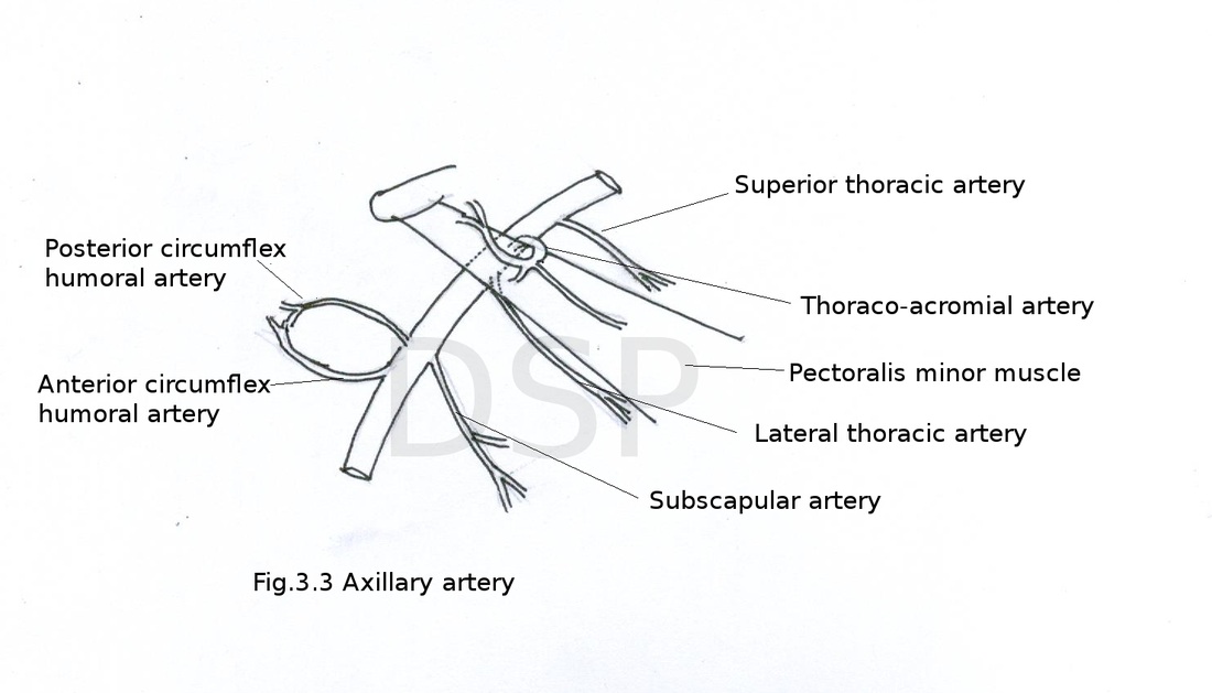
Branches of axillary artery
1 branch from first part, 2 branches from second part and three branches from third part.
1. Superior thoracic artery 2. Thoraco-acromial artery 3. Lateral thoracic artery 4. Subscapular artery 5. Anterior circumflex humeral artery 6. Posterior circumflex humeral artery
1. Superior thoracic artery:
This is 1st branch from 1st part of axillary artery which runs along upper margin of pectoralis minor muscle and supply pectoralis minor and major muscle.
2.Thoraco-acromial artery:
It is the branch from second part gives out 4 branches pectoral, acromial clavicular and deltoid. Pectoral branch supply pectoral muscles, acromial branch forms anastomoses in relation with acromion process, clavicular branch supply subclavius muscle and sterno- clavicular joint, deltoid branch supply deltoid muscle and participate in anastomoses around acromion process.
3.Lateral thoracic artery:
It runs along lower margin of pectoralis minor muscle. It gives out branches to mammary gland in the female.
4. Subscapular artery:
This is a largest branch of third part of axillary artery runs downloads along lower margin of subscapularis muscle. It gives out a large branch circumflex scapular artery and a branch in subscapular fossa. It forms anastomoses around scapula.
5.Anterior circumflex humeral artery:
It is a branch arising from third part of axillary artery. It passes in front of intertubercular sulcus and anastomoses with posterior circumflex humeral artery around surgical neck of humerus. It gives a smallascending branch passing through bicipital groove. Ascending branch supply shoulder joint and humerus.
6. Posterior circumflex humeral artery:
It passes through quadrangular intermuscular space alongwith axillary nerve. It anastomoses with anterior circumflex humeral artery around surgical neck of humerus.It gives out a descnding branch. Descending branch anastomoses with ascending branch of profunda brachii artery.
It is a continuation of third part of subclavian artery. It extends from outer margin of first rib to lower margin of teres major muscle afterwards it continue as brachial artery this artery is divided into 3 parts by pectoralis minor muscle. First part proximal to muscle, second part posterior to muscle and third part distal to the muscle.
Relations of axillary artery
First part
Anterior relation: skin, superficial fascia, platysma and supraclavicular nerves deep fascia pectoralis major muscle, clavipectoral fascia, a communicating loop between lateral and medial pectoral nerve. Axillary artery
Posterior relations: first two digitations of serratus anterior muscle with its nerve, medial cord of brachial plexus and it's medial pectoral branch.
Lateral relations: lateral and posterior cords of brachial plexus.
Medial relations : axillary vein.
Second part
Anterior relations: skin, fascia, pectoralis major, pectoralis minor.
Posterior relations: Subscapularis muscle, posterior cord of brachial plexus.
Lateral relations: lateral cord
Medial relations: medial cord, axillary vein , medial pectoral nerve.
Third part
Anterior relations: skin, fascia, pectoralis major, medial root of median nerve
Posterior relations: subscapularis, latissimis dorsi and teres major muscles axillary nerve and radial nerve.
Lateral relations: coracobrachialis muscle, trunk of median nerve, lateral root of median nerve and musculocutaneous nerve.
Medial relations: axillary vein, medial cutaneous nerve of arm and medial cutaneous nerve of forearm.
Branches of axillary artery
1 branch from first part, 2 branches from second part and three branches from third part.
1. Superior thoracic artery 2. Thoraco-acromial artery 3. Lateral thoracic artery 4. Subscapular artery 5. Anterior circumflex humeral artery 6. Posterior circumflex humeral artery
1. Superior thoracic artery:
This is 1st branch from 1st part of axillary artery which runs along upper margin of pectoralis minor muscle and supply pectoralis minor and major muscle.
2.Thoraco-acromial artery:
It is the branch from second part gives out 4 branches pectoral, acromial clavicular and deltoid. Pectoral branch supply pectoral muscles, acromial branch forms anastomoses in relation with acromion process, clavicular branch supply subclavius muscle and sterno- clavicular joint, deltoid branch supply deltoid muscle and participate in anastomoses around acromion process.
3.Lateral thoracic artery:
It runs along lower margin of pectoralis minor muscle. It gives out branches to mammary gland in the female.
4. Subscapular artery:
This is a largest branch of third part of axillary artery runs downloads along lower margin of subscapularis muscle. It gives out a large branch circumflex scapular artery and a branch in subscapular fossa. It forms anastomoses around scapula.
5.Anterior circumflex humeral artery:
It is a branch arising from third part of axillary artery. It passes in front of intertubercular sulcus and anastomoses with posterior circumflex humeral artery around surgical neck of humerus. It gives a smallascending branch passing through bicipital groove. Ascending branch supply shoulder joint and humerus.
6. Posterior circumflex humeral artery:
It passes through quadrangular intermuscular space alongwith axillary nerve. It anastomoses with anterior circumflex humeral artery around surgical neck of humerus.It gives out a descnding branch. Descending branch anastomoses with ascending branch of profunda brachii artery.
Axillary vein
Basilic vein continue as axillary vein. Axillary vein continue as subclavian vein at first rib. During its course axillary vein lies medial to axillary artery. Cephalic vein opens in axillary vein near its upper part. Tributaries of axillary vein are same as branches of axillary artery. It lies outside axillary sheath to allow expansion during increased venous return.
Applied anatomy: axillary vein thrombosis is a condition in it thrombosis of axillary vein occurs due to continuous pressure on axillary vein by subclavius muscle in painters cause is abducted position of upper limb continuously.
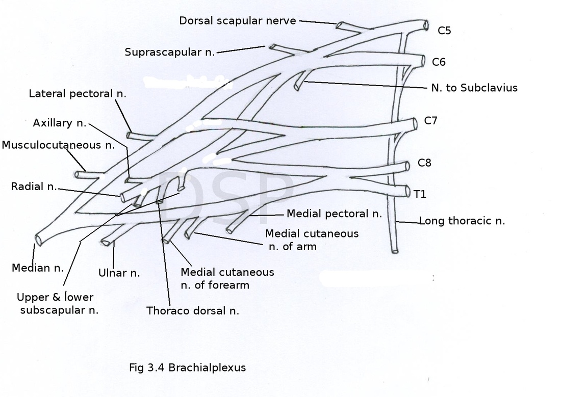
Brachial plexus
Brachial plexus formed by 1. roots 2. trunks 3. divisions 4. cords.
1. Roots : Roots are from ventral rami of cervical nerves C5, C6, C7, C8 and thoracic T1. Sometimes it may be prefixed or postfixed in prefixed type C4 route contribute and in postfixed type T2 root contribute in formation of brachial plexus.
2.Trunks : There are three trunks upper trunk, middle trunk and lower trunk. Formation is as follows upper trunk C5 and C6 roots, middle trunk C7, lower trunk from C8 and T1 roots.
3. Divisions: Trunks divide into anterior division and posterior division.
4. Cords:There are three cords lateral cord, medial cord and posterior cord. Lateral cord formed by anterior divisions of upper and middle trunks, Medial cord by anterior division of lower trunk, posterior cord by posterior divisions of all 3 trunks.
Branches
Branches from roots:
1. Long thoracic nerve C5 C6 C7
2. Dorsal scapular nerve C5
Branches from trunks:
1.Nerve to Subclavius C5 C6
2. Suprascapular nerve C5 C6
Branches from cords:
From Lateral cord
1. Lateral pectoral nerve C5 C6 C7
2. Musculocutaneous nerve C5 C6 C7
3. Lateral root of median nerve
From Medial cord
1. Medial pectoral nerve C8 T1
2. Medial cutaneous nerve of arm C8 T1
3. Medial cutaneous nerve of forearm C8 T1
4. Ulnar nerve C7 C8 T1
5. Medial root of median nerve
From posterior cord
1. Upper subscapular nerve C5 C6
2. Lower subscapular nerve C5 C6
3. Thoracodorsal nerve C6 C7 C8
4. Axillary nerve C5 C6
5. Radial nerve C5 C6 C7 C8 T1
Long thoracic nerve (Nerve to serratus anterior):
It arises from roots C5 C6 and C7 then it goes downwards after piercing scalenus medius muscle enters in axilla to supply serratus anterior muscle.
Dorsal scapular nerve:
It arises from root C5 during its course it pierces scalenus medius muscle then goes downwards deep to levator scapulae muscle and supply rhomboideus major and rhomboideus minor muscle.
Suprascapular nerve:
It arises from roots C5 and C6 of upper trunk goes backward passes through suprascapular foramen to supply supraspinatus, infraspinatus muscles and shoulder joint.
Nerve to subclavius:
It arises from C5 C6 roots of a upper trunk of brachial plexus goes downwards to supply subclavius muscle.
Lateral pectoral nerve:
This nerve after its exit from roots join with medial pectoral nerve and supply pectralis major and pectoralis minor muscles.
Musculocutaneous nerve:
After its exit it runs downwards and laterally lies lateral to third part of axillary artery. Passes between two heads of coracobrachialis again goes downwards in between biceps brachii and brachialis muscles. Then it croses elbow joint on lateral aspect of elbow joint continue downward as lateral cutaneous nerve of forearm and supply skin of anterior and lateral part of forearm up to thenar eminence. During its course it supply coracobrachialis muscle, two heads of biceps brachii and medial part of brachialis muscle also a small branch to elbow joint.
Medial pectoral nerve:
It is a branch of medial cord. After its exit lies posterior to first part of axillary artery then it joins with lateral pectoral nerve. It passes through pectoralis minor muscles. Then supply pectoralis major and pectoralis minor muscle during its course.
Medial cutaneous nerve of arm:
It is a branch of medial cord of brachial plexus. It lies medially goes downwards pierces deep fascia at middle part of arm to supply skin of medial side of the arm below point of piercing.
Medial cutaneous nerve of forearm:
It is a branch of medial cord of brachial plexus. It runs on medial aspect of arm pierces deep fascia of arm in the middle. Divides into two branches anterior and posterior. It supply skin of lower part of arm and medial aspect of forearm up to wrist joint.
Median nerve:
It is formed by lateral root and medial root. Lateral root is from lateral cord and medial root is from medial cord of brachial plexus. Both roots join with each other anterior to third part of axillary artery then goes downwards and laterally.
Ulnar nerve :
It is a branch of medial cord of brachial plexus. After its formation it goes downwards between third part of axillary artery and axillary vein then descends downwards posterior to medial epicondyle of humerus.
Upper subscapular nerve:
It is a branch of posterior cord . It supply upper part of subscapularis muscle,
Lower subscapular nerve:
It is a branch of posterior cord. Supply lower part of subscapularis and teres major muscle.
Thoracodorsal nerve:
It is a branch of posterior cord. During its course it runs downwards along with subscapular artery and supply latissimus dorsi muscle.
Axillary nerve:
It is a branch of posterior cord of brachial plexus . It lies posterior to third part of axillary artery. It passes through quadrangular space and divide into two branches anterior and posterior. Before division it gives out a branch to shoulder joint. Anterior branch passes deep to deltoid muscle in relation with surgical neck of humerus. Posterior branch passes superficial to deltoid finally it continue as cutaneous branch to supply part of skin in relation with deltoid muscle . Anterior branch supply deltoid muscle and skin of anteroinferior part inrelation to deltoid muscle, posterior branch supply teres minor muscle.
Radial nerve:
It is a branch of posterior cord of brachial plexus. It passes downwards laterally and lies posterior to third part of axillary artery. Then go through triangular space lies in relation with shaft of humerus. Branches of radial nerve a branch to long head of triceps, medial head of triceps and posterior cutaneous nerve of arm to supply skin of posterior surface of arm.
Applied anatomy of brachial plexus
Winging of scapula: In this medial border of scapula becomes more prominent when patient try to push with his upper limb. Cause is injury to long thoracic nerve i.e. nerve to serratus anterior. During pushing movement medial margin becomes prominent because of paralysis of serratus anterior muscle. Abduction of upper limb beyond 90 degree is not possible cause is serratus anterior perform this action beyond 90 degree.
Klumpke's paralysis: Claw hand with hyperextension at metacarpophalangeal joint and flexion at interphalangeal joint. Lower trunk of brachial plexus get injured in this. C8 and T1 nerves are involved. Flexor muscles of hand supplied by C8 nerve and muscles of hand supplied by T1 nerve are get paralyzed. Sensory loss along medial margin of forearm and hand occurs. T1 root when involved it causes Horner's syndrome. In horner's syndrome ptosis, miosis, enophthalmos and loss of sweating on the side of lesion i.e. anhydrosis.
Erb's paralysis: When there is injury at erb's point. It causes paralysis in a typical position of upper limb called policeman's tip. Following deformities occur in this arm adducted and medialy rotated, forearm extended at elbow joint and pronated.
Erb's point is a meeting place of 6 nerves 1. root of C5 2. root of C6 3. anterior division 4. posterior division of upper trunk 5. suprascapular nerve 6. nerve to subclavius.
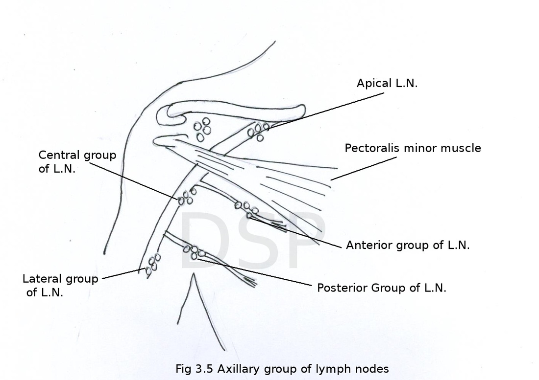
Axillary lymph nodes
These are divided into five groups 1. Anterior 2. Posterior 3. Lateral 4. Central 5. Apical
1. Anterior group: Anterior group of lymph nodes lies in relation with lateral thoracic vessels and it drains from breast mainly then from axillary tail of breast.
2. Posterior group : This lymph nodes lies in relation with subscapular vessels. They drain from dorsal body wall up to iliac crest .
3. Lateral group: Lies along lateral wall of axilla. These drains lymphatics from upper limb.
4. Central group: These are present in axillary fat. Receive lymphatics from other axillary lymph nodes and drains into a apical group of lymph nodes.
5. Apical group: These lymph nodes lies near apex of axilla and drains from upper limb through lymphatic vessels which runs along with cephalic vein, from upper part of breast and other axillary lymph nodes mainly central group of lymph nodes.
Applied anatomy of axilla:
Axillary abscess: Abscess in axilla occurs because of the presence of abundant fat and lymphnodes. Lymphnodes may get infected and suppression of lymphnodes forms abscess in axilla.
Brachial plexus formed by 1. roots 2. trunks 3. divisions 4. cords.
1. Roots : Roots are from ventral rami of cervical nerves C5, C6, C7, C8 and thoracic T1. Sometimes it may be prefixed or postfixed in prefixed type C4 route contribute and in postfixed type T2 root contribute in formation of brachial plexus.
2.Trunks : There are three trunks upper trunk, middle trunk and lower trunk. Formation is as follows upper trunk C5 and C6 roots, middle trunk C7, lower trunk from C8 and T1 roots.
3. Divisions: Trunks divide into anterior division and posterior division.
4. Cords:There are three cords lateral cord, medial cord and posterior cord. Lateral cord formed by anterior divisions of upper and middle trunks, Medial cord by anterior division of lower trunk, posterior cord by posterior divisions of all 3 trunks.
Branches
Branches from roots:
1. Long thoracic nerve C5 C6 C7
2. Dorsal scapular nerve C5
Branches from trunks:
1.Nerve to Subclavius C5 C6
2. Suprascapular nerve C5 C6
Branches from cords:
From Lateral cord
1. Lateral pectoral nerve C5 C6 C7
2. Musculocutaneous nerve C5 C6 C7
3. Lateral root of median nerve
From Medial cord
1. Medial pectoral nerve C8 T1
2. Medial cutaneous nerve of arm C8 T1
3. Medial cutaneous nerve of forearm C8 T1
4. Ulnar nerve C7 C8 T1
5. Medial root of median nerve
From posterior cord
1. Upper subscapular nerve C5 C6
2. Lower subscapular nerve C5 C6
3. Thoracodorsal nerve C6 C7 C8
4. Axillary nerve C5 C6
5. Radial nerve C5 C6 C7 C8 T1
Long thoracic nerve (Nerve to serratus anterior):
It arises from roots C5 C6 and C7 then it goes downwards after piercing scalenus medius muscle enters in axilla to supply serratus anterior muscle.
Dorsal scapular nerve:
It arises from root C5 during its course it pierces scalenus medius muscle then goes downwards deep to levator scapulae muscle and supply rhomboideus major and rhomboideus minor muscle.
Suprascapular nerve:
It arises from roots C5 and C6 of upper trunk goes backward passes through suprascapular foramen to supply supraspinatus, infraspinatus muscles and shoulder joint.
Nerve to subclavius:
It arises from C5 C6 roots of a upper trunk of brachial plexus goes downwards to supply subclavius muscle.
Lateral pectoral nerve:
This nerve after its exit from roots join with medial pectoral nerve and supply pectralis major and pectoralis minor muscles.
Musculocutaneous nerve:
After its exit it runs downwards and laterally lies lateral to third part of axillary artery. Passes between two heads of coracobrachialis again goes downwards in between biceps brachii and brachialis muscles. Then it croses elbow joint on lateral aspect of elbow joint continue downward as lateral cutaneous nerve of forearm and supply skin of anterior and lateral part of forearm up to thenar eminence. During its course it supply coracobrachialis muscle, two heads of biceps brachii and medial part of brachialis muscle also a small branch to elbow joint.
Medial pectoral nerve:
It is a branch of medial cord. After its exit lies posterior to first part of axillary artery then it joins with lateral pectoral nerve. It passes through pectoralis minor muscles. Then supply pectoralis major and pectoralis minor muscle during its course.
Medial cutaneous nerve of arm:
It is a branch of medial cord of brachial plexus. It lies medially goes downwards pierces deep fascia at middle part of arm to supply skin of medial side of the arm below point of piercing.
Medial cutaneous nerve of forearm:
It is a branch of medial cord of brachial plexus. It runs on medial aspect of arm pierces deep fascia of arm in the middle. Divides into two branches anterior and posterior. It supply skin of lower part of arm and medial aspect of forearm up to wrist joint.
Median nerve:
It is formed by lateral root and medial root. Lateral root is from lateral cord and medial root is from medial cord of brachial plexus. Both roots join with each other anterior to third part of axillary artery then goes downwards and laterally.
Ulnar nerve :
It is a branch of medial cord of brachial plexus. After its formation it goes downwards between third part of axillary artery and axillary vein then descends downwards posterior to medial epicondyle of humerus.
Upper subscapular nerve:
It is a branch of posterior cord . It supply upper part of subscapularis muscle,
Lower subscapular nerve:
It is a branch of posterior cord. Supply lower part of subscapularis and teres major muscle.
Thoracodorsal nerve:
It is a branch of posterior cord. During its course it runs downwards along with subscapular artery and supply latissimus dorsi muscle.
Axillary nerve:
It is a branch of posterior cord of brachial plexus . It lies posterior to third part of axillary artery. It passes through quadrangular space and divide into two branches anterior and posterior. Before division it gives out a branch to shoulder joint. Anterior branch passes deep to deltoid muscle in relation with surgical neck of humerus. Posterior branch passes superficial to deltoid finally it continue as cutaneous branch to supply part of skin in relation with deltoid muscle . Anterior branch supply deltoid muscle and skin of anteroinferior part inrelation to deltoid muscle, posterior branch supply teres minor muscle.
Radial nerve:
It is a branch of posterior cord of brachial plexus. It passes downwards laterally and lies posterior to third part of axillary artery. Then go through triangular space lies in relation with shaft of humerus. Branches of radial nerve a branch to long head of triceps, medial head of triceps and posterior cutaneous nerve of arm to supply skin of posterior surface of arm.
Applied anatomy of brachial plexus
Winging of scapula: In this medial border of scapula becomes more prominent when patient try to push with his upper limb. Cause is injury to long thoracic nerve i.e. nerve to serratus anterior. During pushing movement medial margin becomes prominent because of paralysis of serratus anterior muscle. Abduction of upper limb beyond 90 degree is not possible cause is serratus anterior perform this action beyond 90 degree.
Klumpke's paralysis: Claw hand with hyperextension at metacarpophalangeal joint and flexion at interphalangeal joint. Lower trunk of brachial plexus get injured in this. C8 and T1 nerves are involved. Flexor muscles of hand supplied by C8 nerve and muscles of hand supplied by T1 nerve are get paralyzed. Sensory loss along medial margin of forearm and hand occurs. T1 root when involved it causes Horner's syndrome. In horner's syndrome ptosis, miosis, enophthalmos and loss of sweating on the side of lesion i.e. anhydrosis.
Erb's paralysis: When there is injury at erb's point. It causes paralysis in a typical position of upper limb called policeman's tip. Following deformities occur in this arm adducted and medialy rotated, forearm extended at elbow joint and pronated.
Erb's point is a meeting place of 6 nerves 1. root of C5 2. root of C6 3. anterior division 4. posterior division of upper trunk 5. suprascapular nerve 6. nerve to subclavius.
Axillary lymph nodes
These are divided into five groups 1. Anterior 2. Posterior 3. Lateral 4. Central 5. Apical
1. Anterior group: Anterior group of lymph nodes lies in relation with lateral thoracic vessels and it drains from breast mainly then from axillary tail of breast.
2. Posterior group : This lymph nodes lies in relation with subscapular vessels. They drain from dorsal body wall up to iliac crest .
3. Lateral group: Lies along lateral wall of axilla. These drains lymphatics from upper limb.
4. Central group: These are present in axillary fat. Receive lymphatics from other axillary lymph nodes and drains into a apical group of lymph nodes.
5. Apical group: These lymph nodes lies near apex of axilla and drains from upper limb through lymphatic vessels which runs along with cephalic vein, from upper part of breast and other axillary lymph nodes mainly central group of lymph nodes.
Applied anatomy of axilla:
Axillary abscess: Abscess in axilla occurs because of the presence of abundant fat and lymphnodes. Lymphnodes may get infected and suppression of lymphnodes forms abscess in axilla.